Topic 5: Muscular Tissues (Muscular System I) / บทที่ 5: เนื้อเยื่อกล้ามเนื้อ และกลไกการหดตัว (ระบบกล้ามเนื้อ 1)
Comparative histology (skeletal vs cardiac vs smooth), connective tissue sheaths, sarcomere bands, sliding filament model, neuromuscular junction (NMJ), E-C coupling, cross-bridge cycling, and muscle pathology.
เปรียบเทียบมิญชวิทยาของกล้ามเนื้อ 3 ชนิด (กล้ามเนื้อลาย กล้ามเนื้อหัวใจ กล้ามเนื้อเรียบ) เยื่อหุ้มกล้ามเนื้อ แถบในซาร์โคเมียร์ แบบจำลองการเลื่อนของเส้นใย รอยต่อประสาทและกล้ามเนื้อ การจับคู่การกระตุ้นกับการหดตัว และพยาธิสภาพของกล้ามเนื้อ
1. Comparison of 3 Muscle Types / 1. ตารางเปรียบเทียบกล้ามเนื้อ 3 ชนิดอย่างละเอียด
The human body contains three distinct types of muscular tissue:
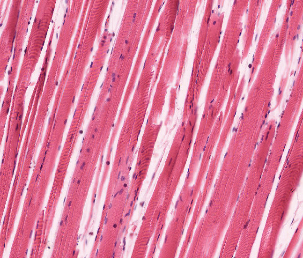
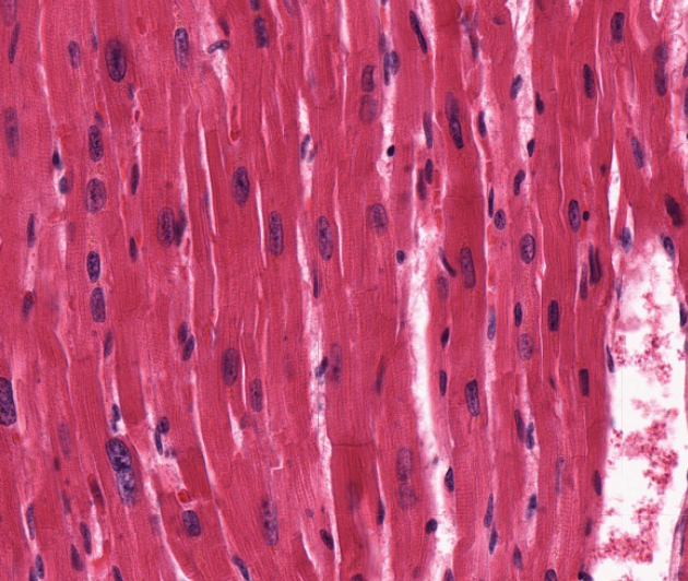
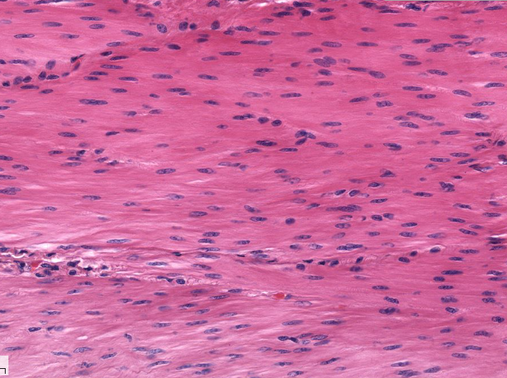
| Feature | Skeletal Muscle | Cardiac Muscle | Smooth Muscle |
|---|---|---|---|
| Location | Attached to skeleton, tongue, pharynx, diaphragm | Heart wall (myocardium) | Walls of hollow visceral organs, vessels, iris, arrector pili |
| Fiber Appearance | Long, unbranched cylinders | Branched cylinders | Fusiform (spindle-shaped) with tapered ends |
| Nuclei | Multinucleated; peripheral beneath sarcolemma | 1 (rarely 2); centrally located | Single, centrally located oval nucleus |
| Striations & Sarcomeres | Yes (prominent A and I bands) | Yes (regular sarcomere organization) | No (non-striated; actin/myosin crisscross) |
| Intercalated Discs | Absent | Present (fascia adherens + desmosomes + gap junctions) | Absent (gap junctions present in single-unit) |
| Control / Regulation | Voluntary (somatic motor neurons) | Involuntary (autonomic nervous system & intrinsic pacemaker) | Involuntary (autonomic NS, hormones, local metabolites) |
| T-Tubules & SR | Triad (1 T-tubule + 2 terminal cisternae at A-I junction) | Diad (1 T-tubule + 1 terminal cisterna at Z disc) | No T-tubules; primitive caveolae; sparse SR |
| Calcium Sensor | Troponin C on thin filament | Troponin C on thin filament | Calmodulin in cytosol (activates MLCK) |
| Calcium Source | Entirely Sarcoplasmic Reticulum (intracellular) | Extracellular ECF (~20%) + SR release (~80%) | Primarily Extracellular ECF + minor SR |
| Regeneration Ability | Limited (via satellite cells) | Essentially None (fibrotic scar repair) | High (hyperplasia via pericytes/mitosis) |
ร่างกายมนุษย์มีเนื้อเยื่อกล้ามเนื้อ 3 ชนิดที่แตกต่างกันชัดเจนทั้งด้านโครงสร้างและหน้าที่:
| ลักษณะสำคัญ | กล้ามเนื้อลาย (Skeletal) | กล้ามเนื้อหัวใจ (Cardiac) | กล้ามเนื้อเรียบ (Smooth) |
|---|---|---|---|
| ตำแหน่งที่พบ | เกาะกับกระดูก ลิ้น กลืน หลอดลม กะบังลม | ผนังหัวใจ (Myocardium) | ผนังอวัยวะภายใน หลอดเลือด ทางเดินอาหาร ม่านตา รูขุมขน |
| รูปร่างเซลล์ | ทรงกระบอกยาว ไม่แตกแขนง | ทรงกระบอกสั้น มีการแตกแขนงเชื่อมกัน | รูปกระสวย (Fusiform) หัวท้ายแหลม |
| จำนวนและตำแหน่งนิวเคลียส | หลายนิวเคลียส (Multinucleated) อยู่ชิดขอบเซลล์ใต้เยื่อหุ้ม | 1 นิวเคลียส (บางครั้ง 2) อยู่ตรงกลางเซลล์ | 1 นิวเคลียสรูปไข่อยู่ตรงกลางเซลล์ |
| ลายตามขวาง (Striations) | มี (เห็นแถบมืด A และแถบสว่าง I ชัดเจน) | มี (จัดเรียงเป็นซาร์โคเมียร์เป็นระเบียบ) | ไม่มีลายตามขวาง (เส้นใยแอกตินและไมโอซินสานเป็นร่างแห) |
| Intercalated Discs | ไม่มี | มี (มี Desmosome ยึดแน่น และ Gap junction ส่งกระแสไฟฟ้า) | ไม่มี (แต่มี Gap junction เชื่อมระหว่างเซลล์) |
| การควบคุม | อำนาจจิตใจ (Voluntary: Somatic motor neuron) | นอกอำนาจจิตใจ (Involuntary: ANS และเซลล์กำหนดจังหวะ) | นอกอำนาจจิตใจ (Involuntary: ANS ฮอร์โมน สารเคมี) |
| T-Tubules และ SR | Triad (1 T-tubule + 2 Terminal cisternae ที่รอยต่อแถบ A-I) | Diad (1 T-tubule กว้าง + 1 Cisterna ที่ระดับ Z disc) | ไม่มี T-tubule มีเพียงหลุม Caveolae และมี SR น้อย |
| โปรตีนตรวจจับ Ca2+ | โทรโปนิน ซี (Troponin C) บนเส้นใยบาง | โทรโปนิน ซี (Troponin C) บนเส้นใยบาง | แคลโมดูลิน (Calmodulin) ในไซโทพลาซึม กระตุ้นเอนไซม์ MLCK |
| แหล่งที่มาของ Ca2+ | จาก Sarcoplasmic Reticulum (SR) ทั้งหมด | จากภายนอกเซลล์ ECF (~20%) + ปล่อยจาก SR (~80%) | ส่วนใหญ่มาจากภายนอกเซลล์ (ECF) ผ่านช่องแคลเซียม |
| การซ่อมแซมและแบ่งตัว | จำกัด (อาศัย Satellite cells ใต้เยื่อเบสเมนต์) | แทบไม่ได้ (ซ่อมแซมด้วยเนื้อเยื่อพังผืด เกิดเป็นรอยแผลเป็น) | แบ่งตัวเพิ่มจำนวนได้ดีมาก (Hyperplasia ผ่าน Pericytes) |
2. Connective Tissue Sheaths & Sarcomere Anatomy / 2. เยื่อหุ้มกล้ามเนื้อ และโครงสร้างซาร์โคเมียร์ (Sarcomere)
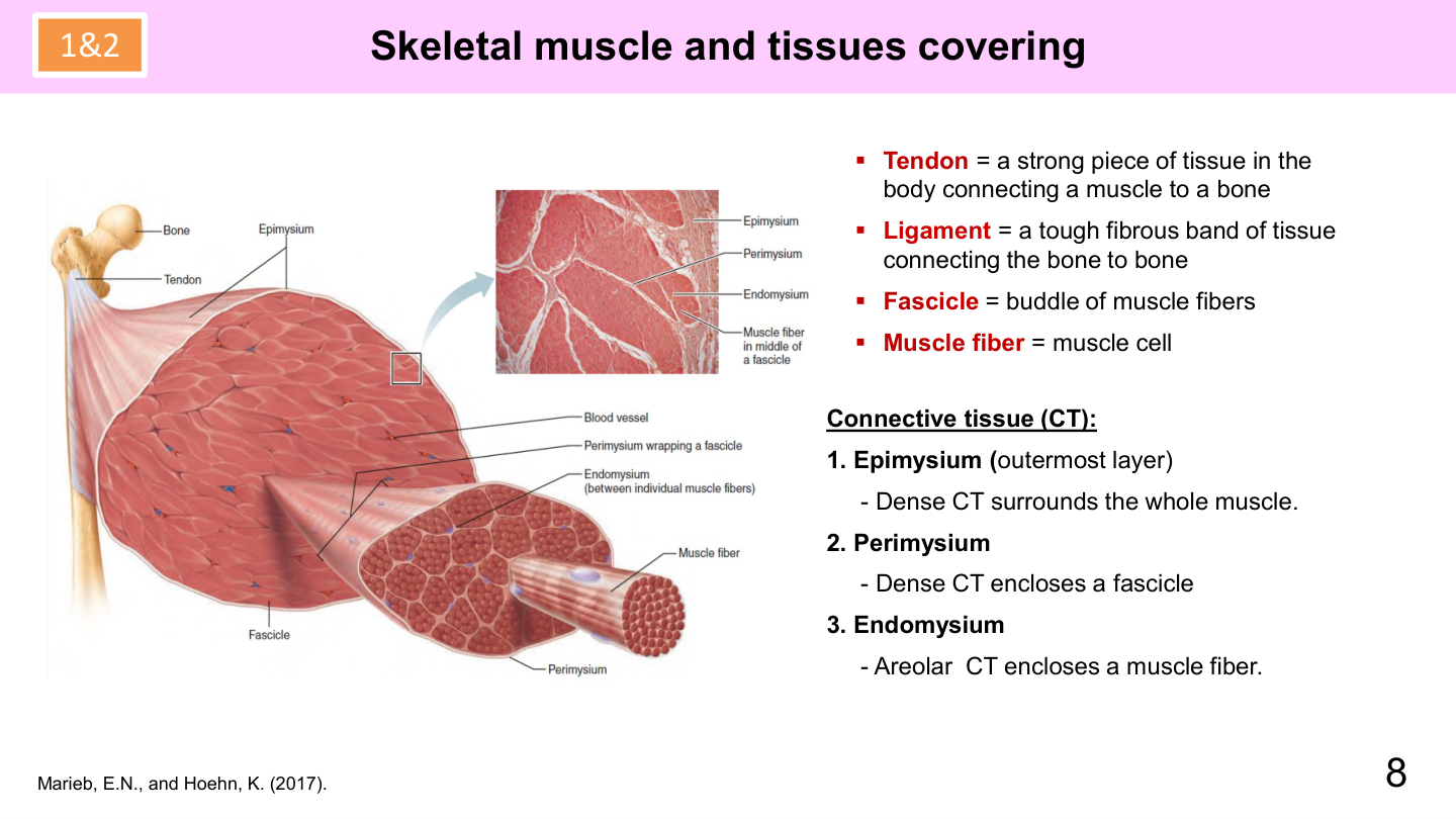
Connective Tissue Coverings
- Epimysium: Dense irregular CT sheath surrounding the entire gross muscle. Continuous with deep fascia.
- Perimysium: Collagenous sheath surrounding bundles of 10–100 muscle fibers called fascicles. Houses neurovascular conduits.
- Endomysium: Delicate layer of areolar CT with reticular fibers wrapping each individual muscle fiber/cell.
- Tendon & Aponeurosis: All three sheaths converge into a rope-like cord (tendon) or broad flat sheet (aponeurosis) anchoring muscle to bone periosteum.
Sarcomere Banding Pattern
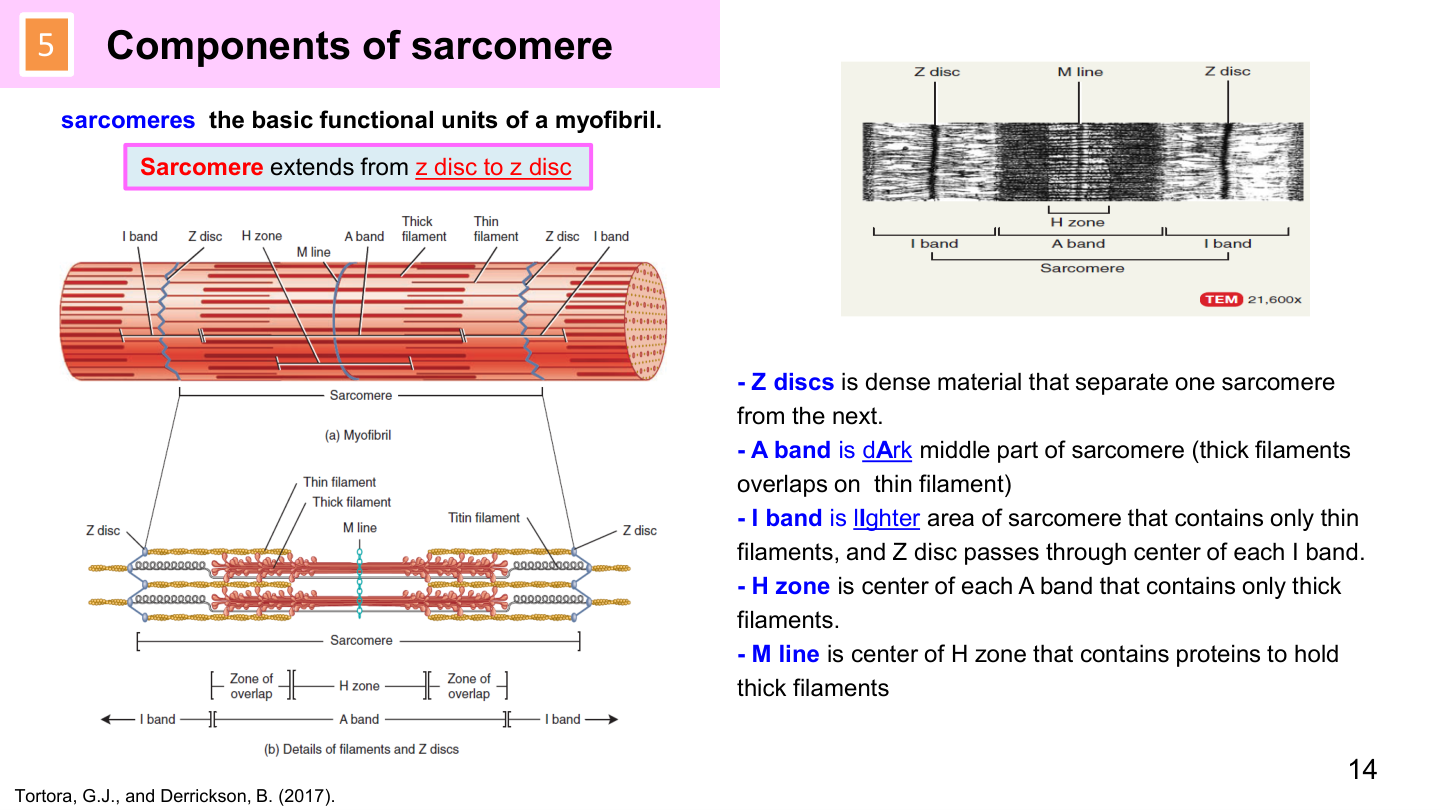
The sarcomere is the functional contractile unit extending between two adjacent Z discs:
| Zone / Band | Description & Molecular Composition | During Contraction (Shortens / Stays Same) |
|---|---|---|
| A Band (Anisotropic) | Dark band spanning entire length of thick (myosin) filaments; includes region of overlap with thin (actin) filaments | STAYS THE SAME LENGTH (myosin filaments do not change length) |
| I Band (Isotropic) | Light band containing ONLY thin (actin) filaments; bisected by the proteinaceous Z disc | SHORTENS / NARROWS (thin filaments pulled toward center) |
| H Zone | Lighter central core within A band containing ONLY thick (myosin) filaments; no actin overlap | SHORTENS AND DISAPPEARS during full contraction |
| M Line | Dark transverse protein line in center of H zone anchoring thick filaments (myomesin) | Stays in center of sarcomere |
| Z Disc (Z Line) | Zigzag protein disc (α-actinin) anchoring thin filaments and titin; defines sarcomere boundaries | DISTANCE BETWEEN Z DISCS SHORTENS (sarcomere shortens) |
เยื่อหุ้มกล้ามเนื้อ 3 ชั้น (Connective Tissue Sheaths)
- Epimysium: ปลอกเนื้อเยื่อเกี่ยวพันหนาหุ้มรอบมัดกล้ามเนื้อทั้งมัด เชื่อมต่อกับ Deep fascia
- Perimysium: ปลอกเนื้อเยื่อเกี่ยวพันหุ้มกลุ่มเส้นใยกล้ามเนื้อ 10-100 เส้น เรียกว่า Fascicle (มัดกล้ามเนื้อย่อย)
- Endomysium: ปลอกเนื้อเยื่อเกี่ยวพันหลวมละเอียด หุ้มล้อมรอบ เส้นใยกล้ามเนื้อเดี่ยวๆ แต่ละเซลล์
- Tendon (เอ็นกล้ามเนื้อ) / Aponeurosis: ปลอกทั้ง 3 ชั้นหลอมรวมที่ปลายกล้ามเนื้อ กลายเป็นเอ็นกลม (Tendon) หรือเอ็นแบน (Aponeurosis) เกาะกับกระดูก
แถบในซาร์โคเมียร์ (Sarcomere Banding Pattern - ออกสอบแล็ปชี้แถบแน่นอน!)
ซาร์โคเมียร์ (Sarcomere): หน่วยทำงานพื้นฐานของการหดตัวของกล้ามเนื้อลาย นับจากแนว Z disc หนึ่งไปยังอีก Z disc หนึ่ง:
| แถบ / โซน | องค์ประกอบทางโมเลกุล | การเปลี่ยนแปลงขณะกล้ามเนื้อหดตัว |
|---|---|---|
| A Band (แถบทึบ) | แถบมืดตามความยาวของเส้นใยหนา (Myosin) ทั้งหมด มีทั้งส่วนที่ซ้อนทับกับ Actin | ความกว้างเท่าเดิมเสมอ (STAYS THE SAME) |
| I Band (แถบสว่าง) | แถบสว่างที่มีเฉพาะ เส้นใยบาง (Actin) เท่านั้น มี Z disc ผ่ากึ่งกลาง | แคบลง / สั้นลง (SHORTENS) |
| H Zone (โซนตรงกลาง) | บริเวณตรงกลางแถบ A ที่มีเฉพาะ เส้นใยหนา (Myosin) เท่านั้น ไม่มี Actin มาซ้อน | แคบลงและหายไป (DISAPPEARS) เมื่อกล้ามเนื้อหดตัวเต็มที่ |
| M Line (เส้นกึ่งกลาง) | เส้นโปรตีนที่ยึดแกนกลางของเส้นใยหนาเข้าด้วยกัน | อยู่ตรงกลางเหมือนเดิม |
| Z Disc (Z Line) | แผ่นโปรตีนหยักรูปซิกแซก ยึดเส้นใยบางและกำหนดขอบเขตของ 1 ซาร์โคเมียร์ | ระยะห่างระหว่าง Z disc หดสั้นเข้าหากัน (ซาร์โคเมียร์หดสั้นลง) |
3. Neuromuscular Junction (NMJ) & 10-Step E-C Coupling / 3. รอยต่อประสานประสาทและกล้ามเนื้อ (NMJ) และ 10 ขั้นตอนการหดตัวของกล้ามเนื้อ
Excitation-Contraction (E-C) coupling converts an electrical neural action potential into mechanical sliding of actin-myosin filaments through 10 sequential steps:
- Action potential reaches the somatic motor axon terminal, depolarizing the presynaptic membrane.
- Voltage-gated $Ca^{2+}$ channels open, allowing $Ca^{2+}$ influx into the terminal bulb.
- $Ca^{2+}$ influx triggers exocytosis of synaptic vesicles, releasing Acetylcholine (ACh) into the 50-nm synaptic cleft.
- ACh diffuses across the cleft and binds to Nicotinic Acetylcholine Receptors (nAChR) on the folded motor end plate.
- Receptor ion channels open ➔ rapid $Na^+$ influx produces local depolarization called the End-Plate Potential (EPP).
- EPP reaches threshold, propagating a muscle action potential along the sarcolemma and deep down into T-tubules.
- Depolarization of T-tubules activates voltage-sensing Dihydropyridine receptors (DHPR), which mechanically uncap Ryanodine receptors (RyR1) on the terminal cisternae of the sarcoplasmic reticulum.
- Massive release of $Ca^{2+}$ surges from the SR lumen into the sarcoplasm (cytosolic $[Ca^{2+}]$ jumps 100-fold).
- $Ca^{2+}$ binds to Troponin C ➔ conformational shift pulls tropomyosin away from the active binding sites on actin.
- Cross-Bridge Cycling:
- Attachment: Cocked myosin head (bound to ADP + Pi) binds actin.
- Power Stroke: Release of Pi and ADP triggers myosin head pivot (~45°), pulling the thin filament toward center of sarcomere.
- Detachment: New molecule of ATP binds to myosin head, causing it to dissociate from actin.
- Reactivation: Myosin ATPase hydrolyzes ATP into ADP + Pi, re-cocking the head into high-energy state.
Muscle Relaxation
Acetylcholinesterase (AChE) destroys ACh in cleft. SERCA pump ($Ca^{2+}$ ATPase) actively pumps cytosolic $Ca^{2+}$ back into SR terminal cisternae. Troponin-tropomyosin complex returns to block actin binding sites.
กลไกการจับคู่การกระตุ้นกับการหดตัว (Excitation-Contraction Coupling) ถ่ายทอดสัญญาณไฟฟ้าจากเซลล์ประสาทสู่การเคลื่อนไหวเชิงกล 10 ขั้นตอน:
- กระแสประสาท (Action potential) เดินทางมาถึงปลายประสาทสั่งการ (Somatic motor axon terminal)
- ช่องแคลเซียมที่เปิดด้วยกระแสไฟฟ้า (Voltage-gated Ca2+ channels) เปิดออก ทำให้ Ca2+ ไหลเข้าสู่ปลายแอกซอน
- Ca2+ กระตุ้นให้ถุงบรรจุสารสื่อประสาทหลอมรวมกับเยื่อหุ้ม ปลดปล่อย Acetylcholine (ACh) สู่ช่องว่างไซแนปส์
- ACh แพร่ข้ามช่องไซแนปส์ไปจับกับตัวรับ Nicotinic ACh Receptor (nAChR) บนรอยพับ Motor end plate
- ช่องไอออนเปิดออก Na+ ไหลทะลักเข้าสู่เซลล์กล้ามเนื้อ เกิดการเปลี่ยนความต่างศักย์เรียกว่า End-Plate Potential (EPP)
- EPP ถึงระดับจุดเริ่ม (Threshold) เกิดกระแสประสาทวิ่งแผ่ไปตามเยื่อหุ้มเซลล์ (Sarcolemma) และวิ่งลึกเข้าสู่ T-tubules
- กระแสไฟฟ้าใน T-tubule กระตุ้นตัวรับ DHPR ซึ่งส่งแรงกลไปดึงเปิดประตูกักเก็บแคลเซียม RyR1 ที่ถุง Terminal cisternae ของ SR
- $Ca^{2+}$ ทะลักออกจาก Sarcoplasmic Reticulum (SR) เข้าสู่ไซโทพลาซึมอย่างรวดเร็ว (ความเข้มข้น Ca2+ พุ่งสูงขึ้น 100 เท่า)
- $Ca^{2+}$ เข้าจับกับโปรตีน Troponin C ทำให้เกิดการเปลี่ยนรูปร่าง ดึงเส้นใย Tropomyosin ให้เปิดออกจากตำแหน่งจับบนสายแอกติน
- วัฏจักรสะพานข้าม (Cross-Bridge Cycle):
- การจับ (Attach): หัวไมโอซินที่มีพลังงานสูง (จับกับ ADP + Pi) เข้าเกาะกับแอกติน
- การดึง (Power stroke): ปลดปล่อย Pi และ ADP หัวไมโอซินหักพับมุม 45 องศา ดึงเส้นใยแอกตินเลื่อนเข้าสู่กึ่งกลางซาร์โคเมียร์
- การหลุด (Detach): โมเลกุล ATP ใหม่เข้ามาจับ ทำให้หัวไมโอซินหลุดออกจากแอกติน
- การตั้งท่าใหม่ (Cocking): เอนไซม์ Myosin ATPase สลาย ATP กลายเป็น ADP + Pi ดึงหัวไมโอซินกลับสู่ท่ายิงพลังงานสูงอีกครั้ง
การคลายตัวของกล้ามเนื้อ (Relaxation)
เอนไซม์ Acetylcholinesterase (AChE) สลายสารสื่อประสาท ACh ในช่องไซแนปส์ และ ปั๊ม SERCA ($Ca^{2+}$ ATPase) ใช้พลังงานสูบ Ca2+ กลับเข้าไปกักเก็บใน SR ส่งผลให้โทรโปนินและโทรโปไมโอซินเลื่อนกลับมาปิดกั้นตำแหน่งจับบนแอกตินตามเดิม
4. Clinical Muscle Pathologies / 4. โรคและพยาธิสภาพของกล้ามเนื้อที่ออกสอบบ่อย
1. Duchenne Muscular Dystrophy (DMD)
X-linked recessive genetic mutation in the dystrophin gene. Dystrophin normally links the actin cytoskeleton to the extracellular basal lamina via the dystrophin-glycoprotein complex. Lack of dystrophin causes sarcolemma tearing during contraction, excess $Ca^{2+}$ influx, muscle fiber necrosis, pseudohypertrophy of calves, and the classic Gower's sign (child climbs up own legs using hands to stand from floor).
2. Myasthenia Gravis (MG)
Autoimmune disorder where autoantibodies attack and destroy nicotinic ACh receptors (nAChR) at the NMJ motor end plate. Causes fluctuating, exertional muscle fatigability: ptosis (drooping eyelid), diplopia (double vision), dysphagia (difficulty swallowing), and dysarthria. Treated with acetylcholinesterase inhibitors (pyridostigmine).
3. Rigor Mortis
Muscular stiffening starting 3–4 hours after death, peaking at 12 hours. Caused by cessation of ATP production. Calcium leaks out of the dying SR and binds troponin, allowing cross-bridges to form. Because NO ATP is available, myosin heads CANNOT detach from actin, trapping muscles in permanent contraction until lysosomes autolyze myofilaments 48–72 hours later.
4. Muscle Regeneration
Adult skeletal muscle fibers are post-mitotic. Repair depends on Satellite Cells (quiescent myogenic stem cells beneath the basal lamina) that proliferate into myoblasts and fuse into multinucleated myotubes. Regenerated mature muscle fibers are histologically identified by CENTRALLY LOCATED NUCLEI (which subsequently migrate to periphery).
1. โรคกล้ามเนื้อเจริญผิดเพี้ยนดูเชนน์ (Duchenne Muscular Dystrophy - DMD)
โรคทางพันธุกรรมแบบ X-linked recessive เกิดจากการกลายพันธุ์ของยีน Dystrophin ซึ่งปกติทำหน้าที่ยึดโยงโครงร่างแอกตินเข้ากับเยื่อหุ้มเซลล์ เมื่อขาดดิสโทรฟิน เยื่อหุ้มเซลล์จะฉีกขาดง่ายเมื่อกล้ามเนื้อหดตัว แคลเซียมไหลทะลักเข้าเซลล์จนกล้ามเนื้อตายและแทนที่ด้วยไขมัน เด็กจะมีกล้ามเนื้อน่องโตเทียม (Pseudohypertrophy) และแสดงอาการ Gower's sign (ใช้มือไต่ขาตัวเองขึ้นมาเพื่อพยุงตัวลุกขึ้นยืนจากพื้น)
2. โรคกล้ามเนื้ออ่อนแรงชนิดร้าย (Myasthenia Gravis - MG)
โรคแพ้ภูมิตัวเองที่สร้างแอนติบอดีมาทำลาย ตัวรับอะเซทิลโคลีน (nAChR) ที่แผ่น Motor end plate ของ NMJ ทำให้ส่งกระแสประสาทสั่งการกล้ามเนื้อได้ลดลงเรื่อยๆ เกิดอาการกล้ามเนื้อล้าง่ายเมื่อใช้งาน เช่น หนังตาตก (Ptosis), มองเห็นภาพซ้อน (Diplopia), กลืนลำบาก, พูดไม่ชัด รักษาด้วยยาต้านเอนไซม์โคลีนเอสเตอเรส (Pyridostigmine)
3. ภาวะศพแข็งทื่อ (Rigor Mortis)
การแข็งเกร็งของกล้ามเนื้อหลังเสียชีวิต 3-4 ชั่วโมง และแข็งสูงสุดที่ 12 ชั่วโมง เกิดจาก การหยุดสร้าง ATP โดยสิ้นเชิง แคลเซียมรั่วออกจาก SR เข้าจับโทรโปนิน ทำให้เกิดสะพานข้ามไมโอซิน-แอกติน แต่เนื่องจากไม่มี ATP ใหม่เข้ามาจับ หัวไมโอซินจึงไม่สามารถหลุดออกจากแอกตินได้ ทำให้กล้ามเนื้อค้างในท่าหดเกร็งถาวร จนกระทั่งเอนไซม์จากไลโซโซมเริ่มย่อยสลายเส้นใยกล้ามเนื้อใน 48-72 ชั่วโมงต่อมา
4. การซ่อมแซมกล้ามเนื้อและเซลล์ดาวเทียม (Satellite Cells)
เซลล์กล้ามเนื้อลายตัวเต็มวัยไม่สามารถแบ่งตัวแบบไมโทซิสได้ การซ่อมแซมอาศัย Satellite Cells (สเต็มเซลล์ที่ซ่อนอยู่ใต้เยื่อเบสเมนต์) ซึ่งจะถูกกระตุ้นให้แบ่งตัวเป็น Myoblast แล้วหลอมรวมเป็น Myotube โดยเส้นใยกล้ามเนื้อที่เพิ่งซ่อมแซมเสร็จใหม่จะมีลักษณะทางมิญชวิทยาจำเพาะคือ มีนิวเคลียสอยู่ตรงกลางเซลล์ (Centrally located nuclei) ก่อนจะเคลื่อนตัวออกไปอยู่ที่ขอบเซลล์ในภายหลัง