Topic 6 of 8
Topic 6: Major Skeletal Muscles (Muscular System II) / บทที่ 6: กล้ามเนื้อโครงร่างมัดสำคัญและกายวิภาคอาจารย์ใหญ่ (ระบบกล้ามเนื้อ 2)
Regional anatomy of major muscles: head, neck, mastication, trunk, abdominal wall, rotator cuff (SITS), upper limb, gluteal region, lower limb, and cadaver pin review checklist.
กายวิภาคของกล้ามเนื้อส่วนต่างๆ: ศีรษะ ใบหน้า การเคี้ยว คอ ลำตัว หน้าท้อง กลุ่มหมุนไหล่ (SITS) แขน ขา สะโพก น่อง และหมายเลขปักเข็มอาจารย์ใหญ่ (Cadaver Pins 1-23)
1. Head, Face & Mastication Muscles / 1. กล้ามเนื้อใบหน้า การแสดงอารมณ์ และการบดเคี้ยว
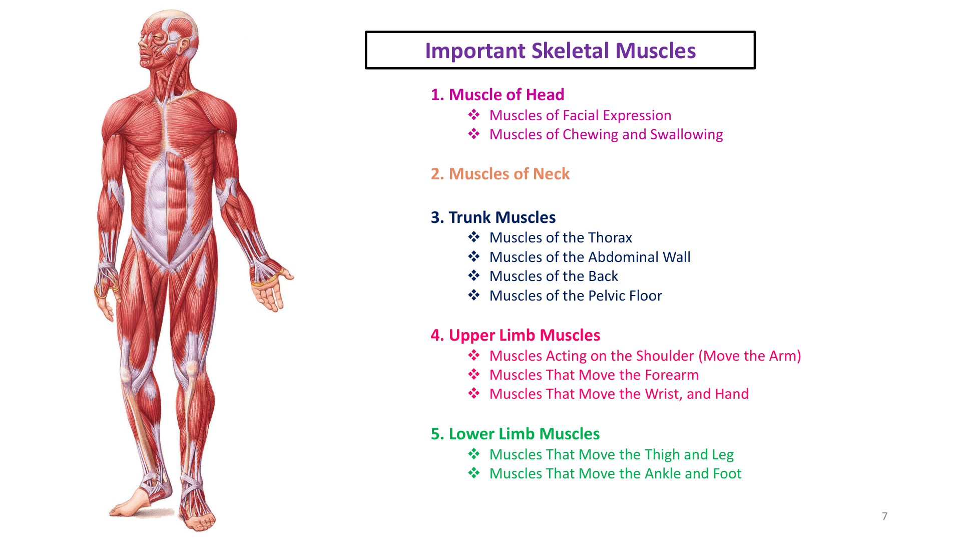
Figure 1: Muscles of Facial Expression (CN VII) and Mastication (CN V3) - Lab Manual Page 52
/
รูปที่ 1: กล้ามเนื้อแสดงสีหน้า (CN VII) และกล้ามเนื้อเคี้ยวอาหาร (CN V3) - คู่มือปฏิบัติการ หน้า 52
Muscles of Facial Expression (Innervated by Facial Nerve - CN VII)
- Frontalis (Frontal belly of Occipitofrontalis): Raises eyebrows, wrinkles forehead in surprise/curiosity.
- Orbicularis oculi: Circular sphincter around eye; closes eyelids tightly (blinking, squinting, sleeping).
- Levator palpebrae superioris: Elevates upper eyelid (innervated by CN III Oculomotor; damage causes ptosis).
- Corrugator supercilii: Draws eyebrows medially and down (frowning, anger, concentration).
- Zygomaticus major & minor: Elevates angle of mouth upward and laterally (smiling and laughing).
- Risorius: Draws corner of mouth straight laterally.
- Buccinator: Compresses cheek against teeth (whistling, blowing trumpet, sucking straw, keeping food on teeth during chewing).
- Orbicularis oris: Oral sphincter; closes, purses, and protrudes lips (kissing, speaking, whistling).
- Mentalis: Wrinkles chin, protrudes lower lip (pouting, doubt).
- Platysma: Superficial thin sheet; tenses skin of neck, depresses mandible and lower lip (horror, fright).
Muscles of Mastication (Innervated by Mandibular Nerve - CN V3)
| Muscle | Origin & Insertion | Action |
|---|---|---|
| Masseter | Zygomatic arch ➔ Ramus and angle of mandible | Elevates mandible (closes jaw); strongest masticatory muscle. |
| Temporalis | Temporal fossa ➔ Coronoid process of mandible | Elevates and retracts mandible (closes jaw and pulls backward). |
| Medial Pterygoid | Pterygoid process & maxilla ➔ Medial surface of mandibular ramus | Elevates mandible, assists in side-to-side grinding chewing. |
| Lateral Pterygoid | Greater wing & lateral pterygoid plate ➔ Mandibular condyle & TMJ capsule | DEPRESSES MANDIBLE (OPENS MOUTH), protracts jaw, side-to-side grinding. (Only masticatory muscle that OPENS jaw). |
กล้ามเนื้อแสดงสีหน้า (ควบคุมโดยเส้นประสาทสมองคู่ที่ 7 - Facial Nerve CN VII)
- Frontalis: ยักคิ้ว ย่นหน้าผากเมื่อตกใจหรือประหลาดใจ
- Orbicularis oculi: กล้ามเนื้อหูรูดรอบดวงตา หลับตา กะพริบตา หรี่ตา
- Levator palpebrae superioris: ยกเปลือกตาบนขึ้น (เลี้ยงโดย CN III Oculomotor หากเป็นอัมพาตจะเกิดหนังตาตก Ptosis)
- Corrugator supercilii: ดึงหัวคิ้วเข้ามาชิดกัน ย่นคิ้วเมื่อโกรธหรือขมวดคิ้ว
- Zygomaticus major & minor: ดึงมุมปากขึ้นบนและไปทางด้านข้าง (กล้ามเนื้อยิ้มและหัวเราะ)
- Risorius: ดึงมุมปากไปด้านข้างตรงๆ
- Buccinator: กล้ามเนื้อกระพุ้งแก้ม กดแก้มแนบฟันขณะเคี้ยว เป่าลม ดูดน้ำจากหลอด เป่าทรัมเป็ต
- Orbicularis oris: กล้ามเนื้อหูรูดรอบริมฝีปาก หุบปาก จู๋ปาก (กล้ามเนื้อจูบ ผิวปาก พูด)
- Mentalis: ดันริมฝีปากล่าง ย่นผิวคางเมื่อทำหน้างอน
- Platysma: แผ่นกล้ามเนื้อบางใต้คางและคอ ดึงคางและมุมปากลงเมื่อตกใจกลัว
กล้ามเนื้อบดเคี้ยวอาหาร (ควบคุมโดยเส้นประสาทสมองคู่ที่ 5 แขนงที่ 3 - Mandibular Nerve CN V3)
| ชื่อกล้ามเนื้อ | จุดเกาะต้นและจุดเกาะปลาย | หน้าที่การทำงาน |
|---|---|---|
| Masseter (แมสซีเตอร์) | Zygomatic arch ➔ กรามล่าง Ramus and angle | ยกขากรรไกรล่างขึ้น (หุบปาก / กัดฟัน) เป็นกล้ามเนื้อเคี้ยวที่ทรงพลังที่สุด |
| Temporalis (เทมพอราลิส) | Temporal fossa ➔ ยอด Coronoid process ของขากรรไกรล่าง | ยกขากรรไกรล่างขึ้นและดึงขากรรไกรถอยหลัง |
| Medial Pterygoid | Pterygoid process ➔ ผิวด้านในของขากรรไกรล่าง | ช่วยยกขากรรไกรล่างขึ้น และบดเคี้ยวเยื้องซ้ายขวา |
| Lateral Pterygoid | Sphenoid ➔ Condyle ของขากรรไกรล่างและข้อต่อ TMJ | กดขากรรไกรล่างลง (อ้าปาก) ยื่นคางไปข้างหน้า เป็นกล้ามเนื้อเคี้ยวมัดเดียวที่ ทำหน้าที่อ้าปาก |
2. Trunk, Thorax & Abdominal Wall Muscles / 2. กล้ามเนื้อลำตัว การหายใจ และผนังหน้าท้อง
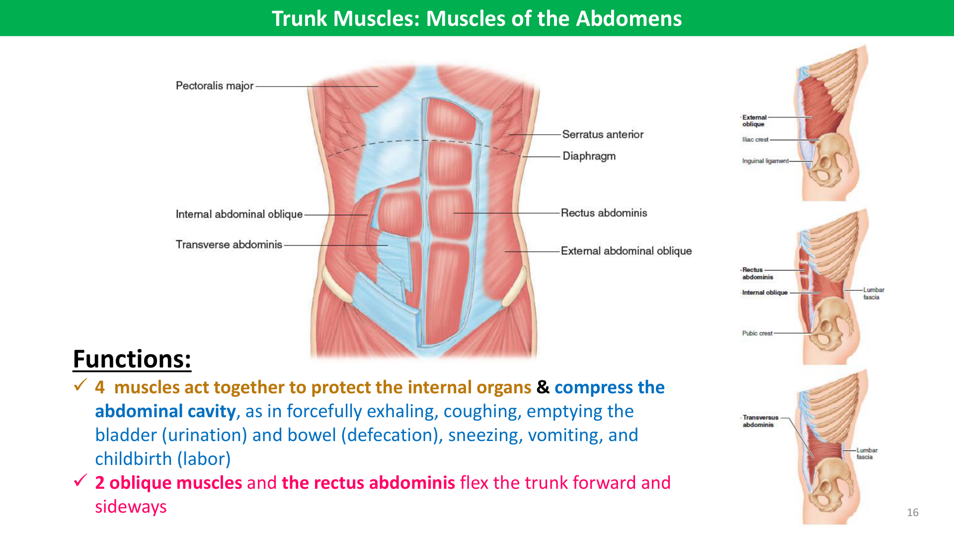
Figure 2: Muscles of the Anterior Thorax and Abdominal Wall (Lab Manual Page 55)
/
รูปที่ 2: กล้ามเนื้อทรวงอกด้านหน้าและผนังหน้าท้อง (คู่มือปฏิบัติการ หน้า 55)
Muscles of Respiration
- Diaphragm: Dome-shaped partition separating thorax and abdomen. Origin: xiphoid process, ribs 7–12, lumbar vertebrae. Insertion: Central tendon of diaphragm. Chief muscle of inspiration (~75% air volume); flattens during contraction. Innervation: Phrenic nerve (C3, C4, C5: "C3, 4, 5 keep the diaphragm alive").
- External Intercostals: Fibers run obliquely down and forward ("hands in pockets"). Elevate ribs during quiet/forced inspiration. Intercostal nerves.
- Internal Intercostals: Fibers run down and backward. Depress ribs during active, forced expiration.
Abdominal Wall Muscles (4 Layers)
- External Abdominal Oblique: Outermost flat muscle; fibers run inferomedially; lower aponeurosis folds to form the inguinal ligament.
- Internal Abdominal Oblique: Middle flat muscle; fibers run superomedially perpendicular to external oblique.
- Transversus Abdominis: Deepest flat muscle; horizontal fibers; compresses abdominal contents.
- Rectus Abdominis: Vertical muscle strap from pubic crest to xiphoid process and costal cartilages 5–7. Segmented by 3 tendinous intersections ("six-pack"); enclosed by rectus sheath; midline fusion forms the fibrous linea alba.
กล้ามเนื้อสำหรับการหายใจ (Respiratory Muscles)
- กะบังลม (Diaphragm): แผ่นกล้ามเนื้อรูปโดมกั้นระหว่างช่องอกและช่องท้อง เกาะที่ Central tendon เป็นกล้ามเนื้อหลักในการหายใจเข้า (75% ของปริมาตรอากาศ) เมื่อหดตัวจะแบนราบลง เพิ่มปริมาตรช่องอก เลี้ยงโดย เส้นประสาทฟรีนิก (Phrenic nerve C3, C4, C5)
- External Intercostals: กล้ามเนื้อระหว่างซี่โครงชั้นนอก ทิศทางเส้นใยเฉียงลงไปข้างหน้า (เหมือนเอามือล้วงกระเป๋า) ทำหน้าที่ยกซี่โครงขึ้นขณะหายใจเข้า
- Internal Intercostals: กล้ามเนื้อระหว่างซี่โครงชั้นใน ทิศทางเส้นใยเฉียงลงไปข้างหลัง กดซี่โครงลงขณะหายใจออกอย่างแรง
กล้ามเนื้อผนังหน้าท้อง 4 ชั้น (Abdominal Wall)
- External Abdominal Oblique: ชั้นนอกสุด เส้นใยเฉียงลงเข้าข้างใน ปลายล่างม้วนเป็นเอ็นขาหนีบ (Inguinal ligament)
- Internal Abdominal Oblique: ชั้นกลาง เส้นใยเฉียงขึ้นเข้าข้างใน ตั้งฉากกับชั้นนอก
- Transversus Abdominis: ชั้นลึกสุด เส้นใยทอดตัวในแนวนอน รัดช่องท้องเพิ่มความดันในช่องท้อง
- Rectus Abdominis: กล้ามเนื้อแถบดิ่งคู่ซ้ายขวา (ซิกแพ็ก) วิ่งจากกระดูกหัวหน่าวขึ้นไปเกาะลิ้นปี่และซี่โครง มีเส้นเอ็นคั่น Tendinous intersections และมีรอยต่อกึ่งกลางหน้าท้องเรียกว่า Linea alba
3. Upper Limb & Rotator Cuff (SITS) / 3. กล้ามเนื้อรยางค์บนและกลุ่มหมุนข้อไหล่ (Rotator Cuff - SITS)
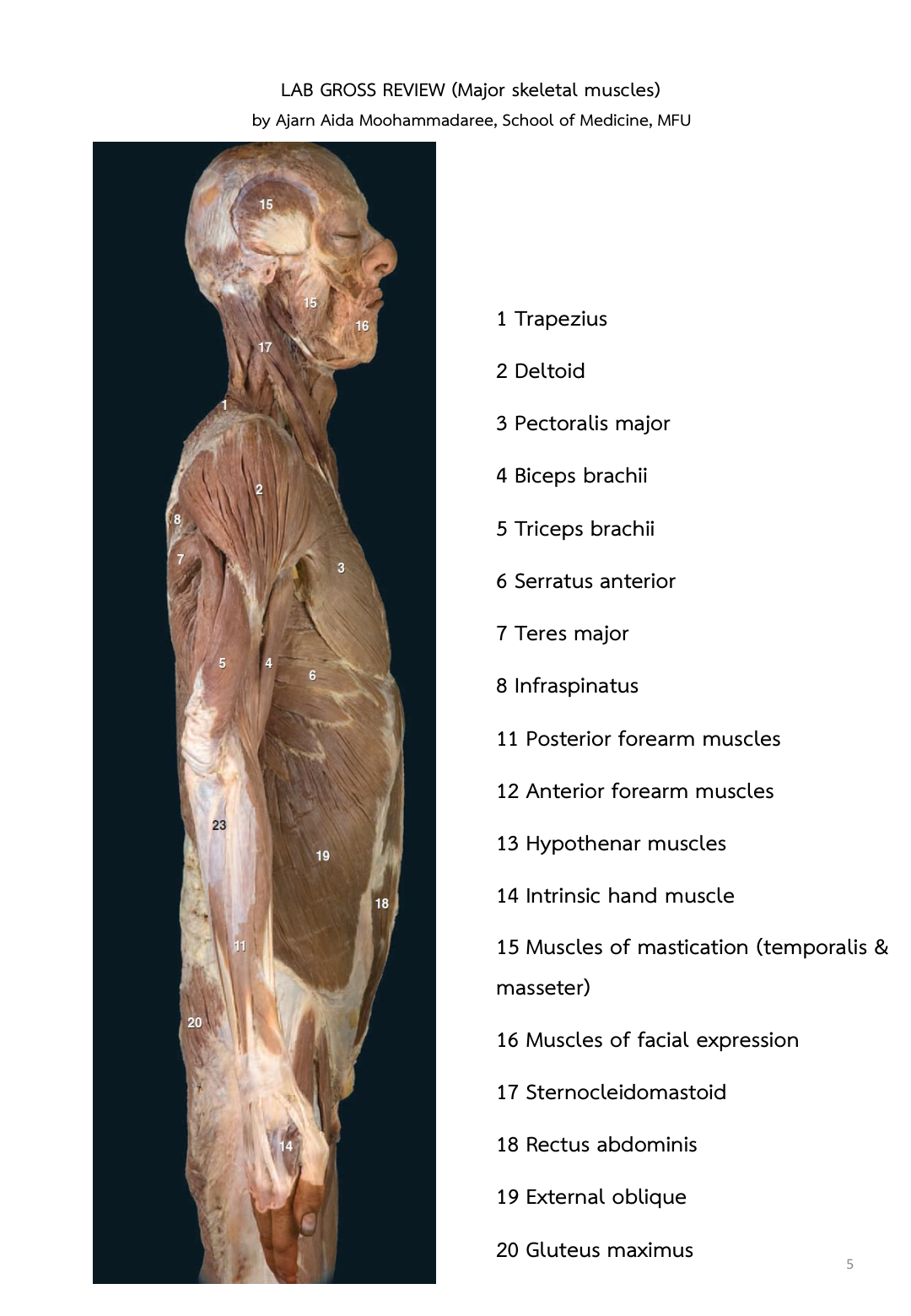
Figure 3: Muscles of the Shoulder and Arm (Deltoid, Biceps brachii, Brachialis, Triceps) - Lab Manual Page 57
/
รูปที่ 3: กล้ามเนื้อหัวไหล่และต้นแขน (Deltoid, Biceps, Brachialis, Triceps) - คู่มือปฏิบัติการ หน้า 57
Rotator Cuff Muscles (SITS) — Crucial Exam Target!
Four scapulohumeral muscles whose tendons encircle and dynamically stabilize the shallow shoulder joint:
| Muscle | Origin & Insertion | Action | Innervation |
|---|---|---|---|
| S - Supraspinatus | Supraspinous fossa ➔ Greater tubercle of humerus (superior facet) | Initiates arm abduction (first 0° to 15°); stabilizes humeral head | Suprascapular nerve (C5, C6) |
| I - Infraspinatus | Infraspinous fossa ➔ Greater tubercle (middle facet) | Lateral (external) rotation of arm | Suprascapular nerve (C5, C6) |
| T - Teres minor | Lateral border of scapula ➔ Greater tubercle (inferior facet) | Lateral (external) rotation and weak adduction | Axillary nerve (C5, C6) |
| S - Subscapularis | Subscapular fossa ➔ Lesser tubercle of humerus | Medial (internal) rotation of arm | Upper & Lower subscapular nerves |
Shoulder & Arm Movers
- Deltoid: Multipennate shoulder cap; middle fibers abduct arm from 15° to 90°; anterior fibers flex and medially rotate; posterior fibers extend and laterally rotate (Axillary nerve).
- Biceps Brachii: Two heads (long from supraglenoid tubercle, short from coracoid process); inserts onto radial tuberosity; powerful supinator of forearm and flexor of elbow when supinated (Musculocutaneous nerve).
- Brachialis: Lies deep to biceps; origin on anterior humerus, insertion on coronoid process of ulna; chief powerful flexor of elbow in all forearm positions (Musculocutaneous nerve).
- Triceps Brachii: 3 heads (long, lateral, medial); inserts on olecranon process of ulna; prime extensor of elbow (Radial nerve).
- Serratus Anterior: Origin on ribs 1-8, inserts on medial scapular border; protracts scapula ("boxer's muscle"). Damage to Long Thoracic Nerve causes Winged Scapula.
กลุ่มกล้ามเนื้อหมุนข้อไหล่ (Rotator Cuff Muscles - ตัวย่อ SITS ที่ออกสอบบ่อยที่สุด!)
กล้ามเนื้อ 4 มัดที่พยุงหัวกระดูกต้นแขนให้เข้าที่ในเบ้าหัวไหล่:
| ชื่อกล้ามเนื้อ | จุดเกาะต้น ➔ จุดเกาะปลาย | หน้าที่การทำงาน | เส้นประสาทควบคุม |
|---|---|---|---|
| S - Supraspinatus | เหนือสันสะบัก ➔ ปุ่ม Greater tubercle ของกระดูกต้นแขน | เริ่มกางแขน 15 องศาแรก (0°-15°) | Suprascapular nerve |
| I - Infraspinatus | ใต้สันสะบัก ➔ ปุ่ม Greater tubercle | หมุนแขนออกด้านนอก (Lateral rotation) | Suprascapular nerve |
| T - Teres minor | ขอบข้างของสะบัก ➔ ปุ่ม Greater tubercle | หมุนแขนออกด้านนอก (Lateral rotation) | Axillary nerve |
| S - Subscapularis | แอ่งด้านหน้าของสะบัก ➔ ปุ่ม Lesser tubercle | หมุนแขนเข้าด้านใน (Medial rotation) | Subscapular nerves |
กล้ามเนื้อสำคัญของหัวไหล่และแขน
- Deltoid (กล้ามเนื้อเดลทอยด์): รูปสามเหลี่ยมคลุมหัวไหล่; เส้นใยส่วนกลาง กางแขนจาก 15° ถึง 90° (เลี้ยงโดย Axillary nerve) เป็นตำแหน่งฉีดยาเข้ากล้ามเนื้อ (IM injection) ที่พบบ่อย
- Biceps Brachii: มี 2 หัว เกาะที่ปุ่มเรเดียล หงายมือ (Supination) และงอข้อศอก (เลี้ยงโดย Musculocutaneous nerve)
- Brachialis: อยู่ลึกใต้ไบเซปส์ เกาะที่กระดูกอัลนา เป็น กล้ามเนื้องอข้อศอกที่ทรงพลังที่สุดในทุกตำแหน่งแขน (เลี้ยงโดย Musculocutaneous nerve)
- Triceps Brachii: มี 3 หัว เกาะที่ปุ่มข้อศอก Olecranon เป็น กล้ามเนื้อเหยียดข้อศอกหลัก (เลี้ยงโดย Radial nerve)
- Serratus Anterior: ยึดสะบักแนบกับผนังช่องอก ยื่นหมัดไปข้างหน้า (Boxer's muscle) หากเส้นประสาท Long Thoracic Nerve เสียหาย จะเกิดภาวะ สะบักลอย (Winged Scapula)
4. Lower Limb Muscles & Clinical Applications / 4. กล้ามเนื้อรยางค์ล่าง จุดฉีดยา และการประยุกต์ทางคลินิก
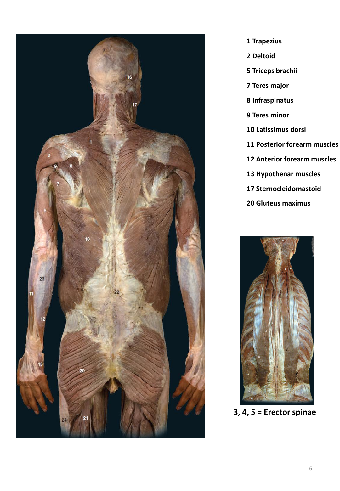
Figure 4: Muscles of the Thigh (Quadriceps, Sartorius, Gracilis, Hamstrings) - Lab Manual Page 61
/
รูปที่ 4: กล้ามเนื้อต้นขา (Quadriceps, Sartorius, Hamstrings) - คู่มือปฏิบัติการ หน้า 61
Gluteal & Thigh Muscle Groups
- Gluteus Maximus: Largest and heaviest; powerful extensor and lateral rotator of thigh at hip (climbing stairs, rising from seated position, running). Inferior gluteal nerve.
- Gluteus Medius & Minimus: Abducts and medially rotates thigh; stabilizes pelvis preventing pelvic tilt during walking. Superior gluteal nerve. Weakness produces positive Trendelenburg sign. Major safe site for intramuscular (IM) ventrogluteal injections (avoids the sciatic nerve).
- Iliopsoas (Psoas major + Iliacus): Inserts onto lesser trochanter of femur; prime, most powerful flexor of thigh at hip.
- Sartorius: Longest strap-like muscle crossing anterior thigh; flexes, abducts, and laterally rotates hip and flexes knee ("cross-legged tailor sitting position").
- Quadriceps Femoris (4 heads): Rectus femoris (crosses hip and knee; flexes hip, extends knee), Vastus lateralis, Vastus medialis, Vastus intermedius. Insert via patellar tendon/ligament onto tibial tuberosity. Prime extensor of knee. Femoral nerve.
- Hamstrings (Posterior Thigh): Biceps femoris, Semitendinosus, Semimembranosus. Origin on ischial tuberosity; flex knee and extend hip. Sciatic nerve (Tibial division).
Leg Muscles (Crus)
- Tibialis Anterior: Anterior compartment; deep fibular nerve; dorsiflexes and inverts foot. Damage to common fibular nerve causes Foot Drop.
- Gastrocnemius & Soleus (Triceps Surae): Posterior compartment; insert via Achilles (calcaneal) tendon onto calcaneus; powerful plantarflexion of foot at ankle (walking, jumping, standing on tiptoes). Tibial nerve.
กล้ามเนื้อสะโพกและต้นขา
- Gluteus Maximus: กล้ามเนื้อมัดใหญ่ที่สุด เหยียดและหมุนข้อสะโพกออกด้านนอกอย่างทรงพลัง (ใช้ขณะวิ่ง ก้าวขึ้นบันได ลุกจากเก้าอี้) เลี้ยงโดย Inferior gluteal nerve
- Gluteus Medius & Minimus: กางขาและหมุนขาเข้าด้านใน พยุงกระดูกเชิงกรานให้สมดุลขณะเดินข้างเดียว หากอ่อนแรงจะเกิด Trendelenburg sign (สะโพกเอียงตก) และบริเวณนี้เป็นตำแหน่ง ฉีดยาเข้ากล้ามเนื้อที่ปลอดภัยที่สุด (Ventrogluteal site) เพราะหลีกเลี่ยงเส้นประสาท Sciatic nerve ขนาดใหญ่
- Iliopsoas (Psoas major + Iliacus): เกาะที่ปุ่ม Lesser trochanter ของกระดูกต้นขา เป็น กล้ามเนื้องอข้อสะโพกที่ทรงพลังที่สุด
- Sartorius: กล้ามเนื้อแถบยาวที่สุดในร่างกาย พาดเฉียงหน้าต้นขา งอ กาง และหมุนสะโพกออกด้านนอก พร้อมงอเข่า (ท่านั่งขัดสมาธิ)
- Quadriceps Femoris (4 หัว): Rectus femoris, Vastus lateralis, Vastus medialis, Vastus intermedius รวมเป็นเอ็นลูกสะบ้าเกาะที่ปุ่มหน้าแข้ง เป็น กล้ามเนื้อเหยียดข้อเข่าหลัก เลี้ยงโดย Femoral nerve
- Hamstrings (กล้ามเนื้อหลังต้นขา 3 มัด): Biceps femoris, Semitendinosus, Semimembranosus มีจุดเกาะต้นที่ปุ่มกระดูกนั่ง (Ischial tuberosity) ทำหน้าที่ งอเข่าและเหยียดสะโพก เลี้ยงโดยเส้นประสาทไซอาติก (Sciatic nerve)
กล้ามเนื้อขาส่วนล่าง (หน้าแข้งและน่อง)
- Tibialis Anterior: หน้าแข้ง เลี้ยงโดย Deep fibular nerve ทำหน้าที่ กระดกข้อเท้าขึ้น (Dorsiflexion) และบิดฝ่าเท้าเข้าใน (Inversion) หากเส้นประสาทบาดเจ็บจะเกิดอาการ ปลายเท้าตก (Foot Drop) เดินลากปลายเท้า
- Gastrocnemius & Soleus (กล้ามเนื้อน่อง): รวมเป็น เอ็นร้อยหวาย (Achilles tendon) เกาะที่กระดูกส้นเท้า Calcaneus ทำหน้าที่ ถีบปลายเท้าลง (Plantarflexion) ใช้ในการเดิน วิ่ง และเขย่งปลายเท้า เลี้ยงโดย Tibial nerve
5. Cadaver Dissection Checklist (Pins 1–23) / 5. หมายเลขปักเข็มอาจารย์ใหญ่ที่ออกสอบแล็ป (Cadaver Pins 1–23)
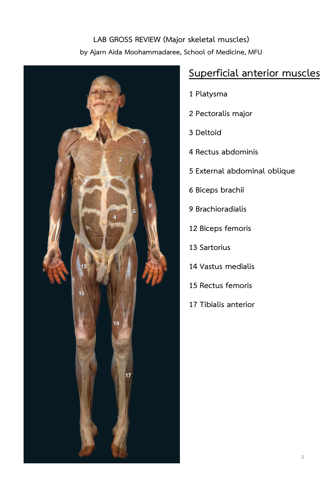
Figure 5: Cadaver Dissection Review - Anterior Muscles (Checklist Review Page 3)
/
รูปที่ 5: กายวิภาคอาจารย์ใหญ่ด้านหน้าพร้อมหมายเลขปักเข็ม (Checklist หน้า 3)
Memorize these official numbered pin locations directly from the course dissection review guide:
| Pin # | Muscle / Structure | Region & Key Identifying Feature |
|---|---|---|
| 1 | Platysma | Superficial neck thin muscle sheet |
| 2 | Pectoralis major | Anterior chest large fan-shaped muscle |
| 3 | Deltoid | Rounded shoulder cap muscle |
| 4 | Rectus abdominis | Vertical anterior abdominal muscle (six-pack) |
| 5 | External abdominal oblique | Lateral abdominal wall with inferomedial fibers |
| 6 | Biceps brachii | Anterior arm muscle with two heads |
| 7 | Triceps brachii | Posterior arm three-headed extensor |
| 8 | Trapezius | Large diamond-shaped superficial back muscle |
| 9 | Brachioradialis | Lateral superficial forearm muscle |
| 10 | Latissimus dorsi | Broad lower back swimmer's muscle |
| 11 | Gluteus maximus | Large superficial buttock muscle |
| 12 | Biceps femoris | Lateral hamstring muscle behind thigh |
| 13 | Sartorius | Long diagonal ribbon crossing anterior thigh |
| 14 | Vastus medialis | Teardrop medial muscle of quadriceps |
| 15 | Rectus femoris | Central superficial muscle of quadriceps |
| 16 | Adductor magnus / group | Medial compartment of thigh |
| 17 | Tibialis anterior | Anterior shin muscle next to tibia crest |
| 18 | Gastrocnemius | Two-bellied superficial calf muscle |
| 19 | Fibularis (peroneus) longus | Lateral compartment of leg |
| 21 | Iliotibial (IT) tract | Thick lateral fascial band of thigh |
| 23 | Quadriceps femoris tendon | Common tendon attaching into patella |
หมายเลขตำแหน่งปักเข็มอาจารย์ใหญ่ตามคู่มือทบทวนแล็ปทางการที่ต้องจำให้แม่น:
| หมายเลขปักเข็ม | ชื่อกล้ามเนื้อ / โครงสร้าง | บริเวณและลักษณะเด่นทางกายวิภาค |
|---|---|---|
| 1 | Platysma | แผ่นกล้ามเนื้อบางใต้ผิวหนังบริเวณคอ |
| 2 | Pectoralis major | กล้ามเนื้ออกมัดใหญ่รูปพัด |
| 3 | Deltoid | กล้ามเนื้อหัวไหล่ทรงสามเหลี่ยม |
| 4 | Rectus abdominis | กล้ามเนื้อหน้าท้องแนวดิ่ง (ซิกแพ็ก) |
| 5 | External abdominal oblique | ผนังหน้าท้องด้านข้าง เส้นใยเฉียงลง |
| 6 | Biceps brachii | กล้ามเนื้อด้านหน้าต้นแขน |
| 7 | Triceps brachii | กล้ามเนื้อด้านหลังต้นแขน (3 หัว) |
| 8 | Trapezius | กล้ามเนื้อสะพายแร่งรูปสี่เหลี่ยมข้าวหลามตัดที่หลังส่วนบน |
| 9 | Brachioradialis | กล้ามเนื้อปลายแขนด้านนอกเกาะจากต้นแขน |
| 10 | Latissimus dorsi | กล้ามเนื้อปีกหลังมัดกว้าง |
| 11 | Gluteus maximus | กล้ามเนื้อแก้มก้นมัดใหญ่ชั้นตื้น |
| 12 | Biceps femoris | กล้ามเนื้อหลังต้นขาด้านนอก (แฮมสตริง) |
| 13 | Sartorius | กล้ามเนื้อแถบยาวพาดเฉียงหน้าต้นขา |
| 14 | Vastus medialis | กล้ามเนื้อต้นขาด้านในรูปหยดน้ำ |
| 15 | Rectus femoris | กล้ามเนื้อตรงกลางด้านหน้าต้นขาชั้นตื้น |
| 16 | Adductor magnus / group | กลุ่มกล้ามเนื้อหุบขาด้านในของต้นขา |
| 17 | Tibialis anterior | กล้ามเนื้อหน้าแข้งแนบชิดกระดูกทีเบีย |
| 18 | Gastrocnemius | กล้ามเนื้อน่องสองหัวชั้นตื้น |
| 19 | Fibularis longus | กล้ามเนื้อขาส่วนล่างด้านข้าง |
| 21 | Iliotibial (IT) tract | แถบเอ็นพังผืดหนาด้านข้างต้นขา |
| 23 | Quadriceps tendon | เอ็นรวมของกล้ามเนื้อควอดริเซพส์ก่อนถึงลูกสะบ้า |
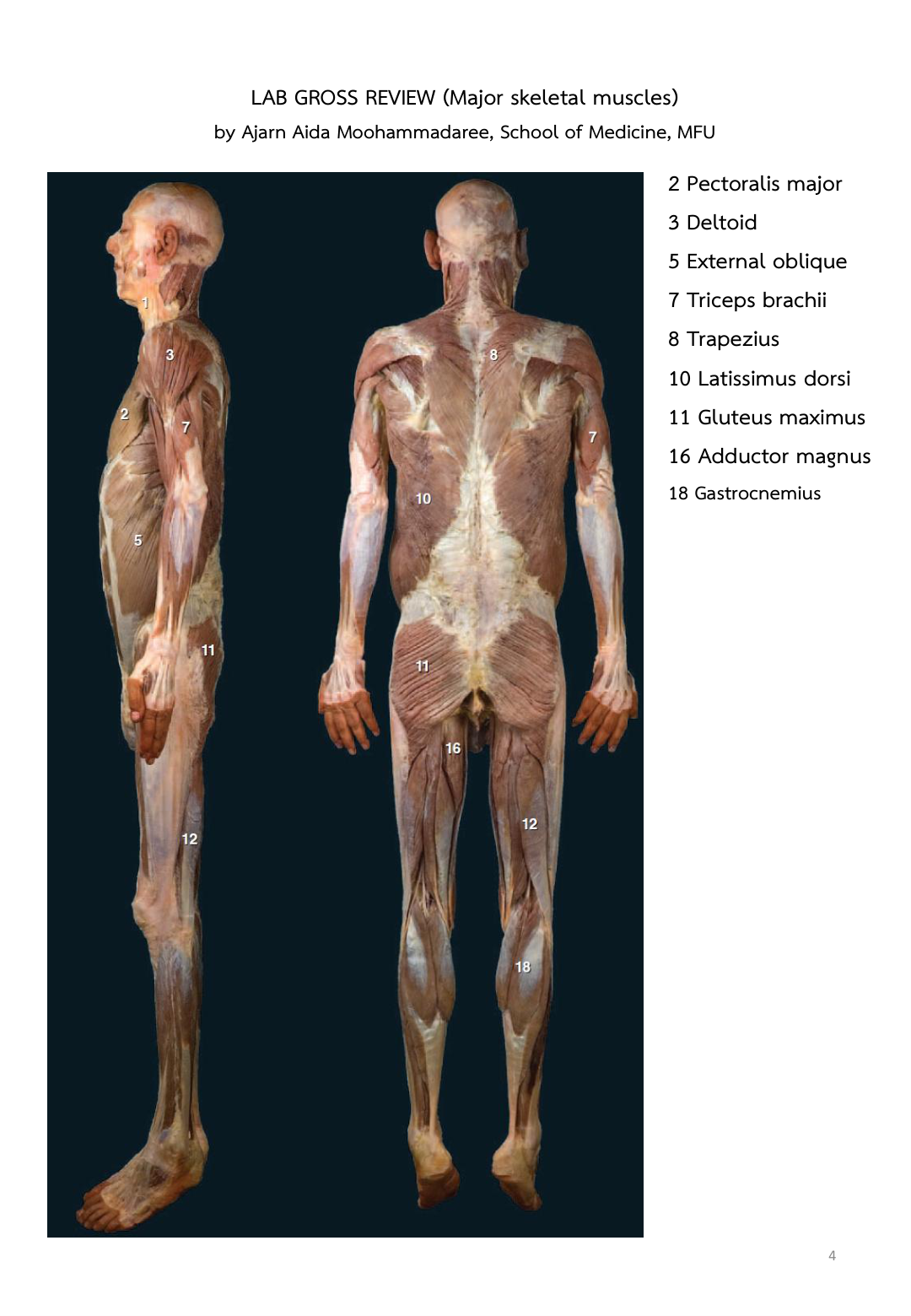
Figure 6: Cadaver Dissection Review - Posterior Muscles (Checklist Review Page 4)
/
รูปที่ 6: กายวิภาคอาจารย์ใหญ่ด้านหลังพร้อมหมายเลขปักเข็ม (Checklist หน้า 4)