Topic 3: Basic Tissues & Integumentary System / บทที่ 3: เนื้อเยื่อพื้นฐานและระบบผิวหนัง
The 4 primary tissue types, cell junctions, epithelial and connective tissue classifications, skin layers, dermal circulation, cutaneous receptors, appendages, burns, and wound healing.
เนื้อเยื่อ 4 ชนิดหลัก รอยต่อระหว่างเซลล์ การจำแนกเนื้อเยื่อบุผิวและเนื้อเยื่อเกี่ยวพัน ชั้นของผิวหนัง ระบบไหลเวียนโลหิต ตัวรับความรู้สึก อวัยวะอนุพันธ์ แผลไฟไหม้ และกระบวนการสมานแผล
1. The 4 Primary Tissues & Cell Junctions / 1. เนื้อเยื่อพื้นฐาน 4 ชนิด และรอยต่อระหว่างเซลล์ (Cell Junctions)
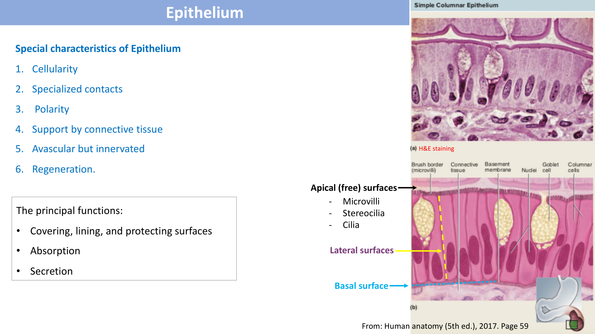
The human body is composed of four primary tissue classes:
- Epithelial Tissue: Avascular, tightly packed sheets covering surfaces and forming glands. Protects, secretes, absorbs, and filters.
- Connective Tissue: Widely separated cells within an abundant extracellular matrix (fibers + ground substance). Binds, supports, protects, and insulates.
- Muscular Tissue: Excitable and contractile cells specialized for mechanical force generation and movement.
- Nervous Tissue: Neurons (impulse generation/conduction) and neuroglia (supporting cells) for sensation, integration, and control.
Cell Junctions
| Junction Type | Major Proteins | Key Function & Typical Location |
|---|---|---|
| Tight Junction (Zonula Occludens) | Occludin, Claudin | Seals adjacent plasma membranes; prevents paracellular fluid leakage (Blood-brain barrier, intestinal lining, stomach). |
| Adherens Junction (Zonula Adherens) | Cadherin + Actin microfilaments | Anchoring belt linking actin cytoskeleton; reinforces apical junctional complexes. |
| Desmosome (Macula Adherens) | Cadherin (Desmoglein/Desmocollin) + Keratin intermediate filaments | "Spot weld" resisting intense mechanical shear/pulling forces (Epidermis of skin, cardiac muscle). |
| Hemidesmosome | Integrin + Keratin | Anchors basal surface of epithelial cell to the underlying basal lamina/basement membrane. |
| Gap Junction (Nexus) | Connexin (forms 6-subunit connexon channel) | Direct cytoplasmic diffusion of small ions, cAMP, metabolites (<1 kDa); electrical coupling (Intercalated discs of cardiac muscle, single-unit smooth muscle). |
ร่างกายมนุษย์ประกอบด้วยเนื้อเยื่อพื้นฐาน 4 ชนิดหลัก:
- เนื้อเยื่อบุผิว (Epithelial Tissue): เซลล์เรียงตัวชิดกันมาก ไม่มีหลอดเลือดมาเลี้ยง (Avascular) คลุมผิวหน้าและสร้างต่อม ทำหน้าที่ป้องกัน ดูดซึม และหลั่งสาร
- เนื้อเยื่อเกี่ยวพัน (Connective Tissue): เซลล์อยู่ห่างกัน ล้อมรอบด้วยสารเคลือบเซลล์ (Extracellular Matrix: เส้นใย + Ground substance) ทำหน้าที่ค้ำจุน ยึดเหนี่ยว ป้องกัน และเป็นทางผ่านของหลอดเลือด
- เนื้อเยื่อกล้ามเนื้อ (Muscular Tissue): เซลล์หดตัวได้ สร้างแรงกลและการเคลื่อนไหว
- เนื้อเยื่อประสาท (Nervous Tissue): เซลล์ประสาท (Neuron) และเซลล์ค้ำจุน (Neuroglia) ทำหน้าที่รับความรู้สึก ประมวลผล และสั่งการ
รอยต่อระหว่างเซลล์ (Cell Junctions)
| ชนิดของรอยต่อ | โปรตีนสำคัญ | หน้าที่และตำแหน่งที่พบ |
|---|---|---|
| Tight Junction (Zonula Occludens) | Occludin, Claudin | ผนึกช่องว่างระหว่างเซลล์ ป้องกันสารรั่วซึมผ่านร่องเซลล์ (เยื่อบุทางเดินอาหาร, Blood-brain barrier) |
| Adherens Junction | Cadherin + เส้นใยแอกติน (Actin) | แถบเชื่อมยึดโครงร่างแอกตินของเซลล์ข้างเคียง |
| Desmosome (Macula Adherens) | Cadherin + เส้นใยเคราติน (Keratin) | "รอยเชื่อมหมุดย้ำ" ทนทานต่อแรงดึงและแรงเฉือนได้สูงมาก (ชั้นหนังกำพร้า, กล้ามเนื้อหัวใจ) |
| Hemidesmosome | Integrin + Keratin | ยึดฐานของเซลล์เยื่อบุผิวเข้ากับแผ่นเยื่อเบสเมนต์ (Basement membrane) |
| Gap Junction (Nexus) | Connexin (รวมเป็นช่อง Connexon) | ช่องทางติดต่อสื่อสารระหว่างไซโทพลาซึมของเซลล์ข้างเคียง ยอมให้ไอออนและสารขนาดเล็กผ่านได้ ถ่ายทอดกระแสประสาทและไฟฟ้าอย่างรวดเร็ว (Intercalated disc ในกล้ามเนื้อหัวใจ) |
2. Epithelial Tissue Classification / 2. การจำแนกชนิดของเนื้อเยื่อบุผิว
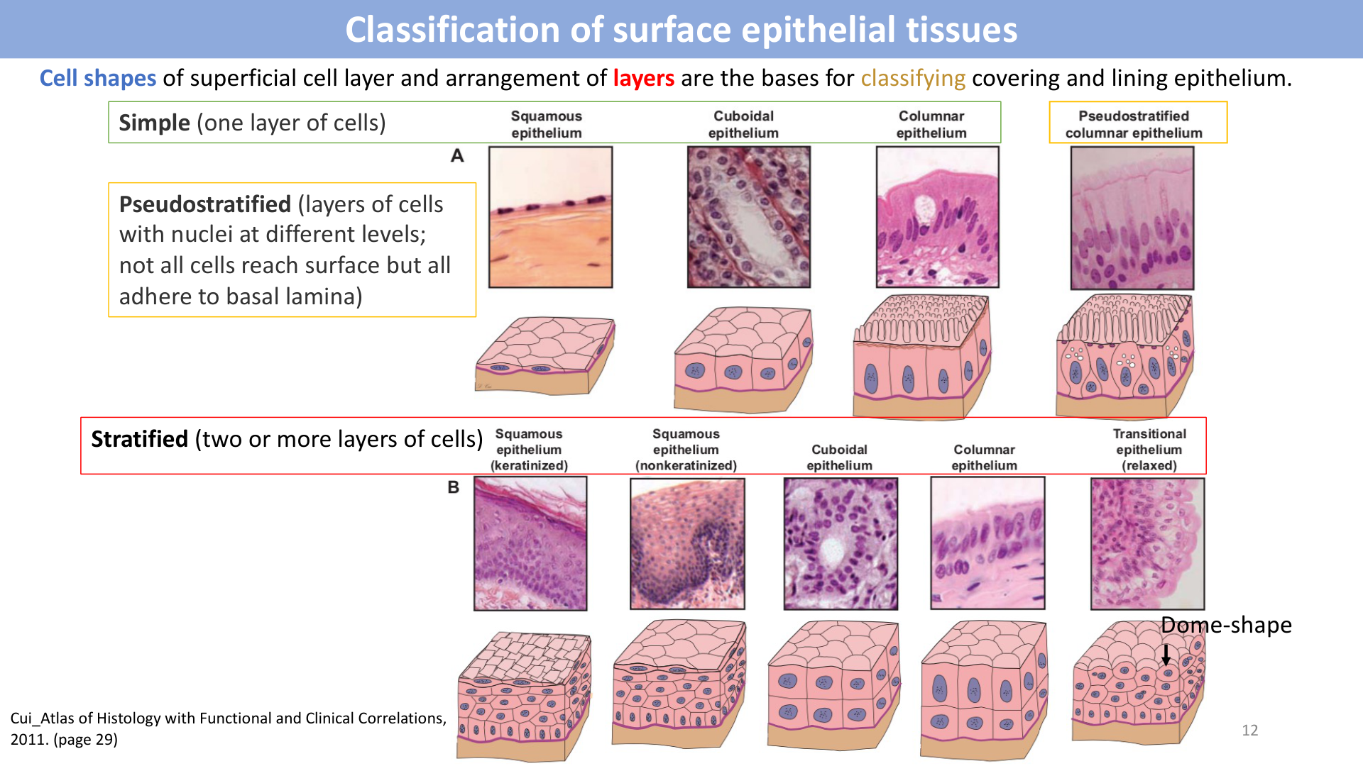
Epithelia are classified by the number of cell layers (Simple, Stratified, Pseudostratified) and the shape of cells at the apical surface (Squamous, Cuboidal, Columnar, Transitional):
| Epithelium Type | Histological Description | Key Locations & Functions |
|---|---|---|
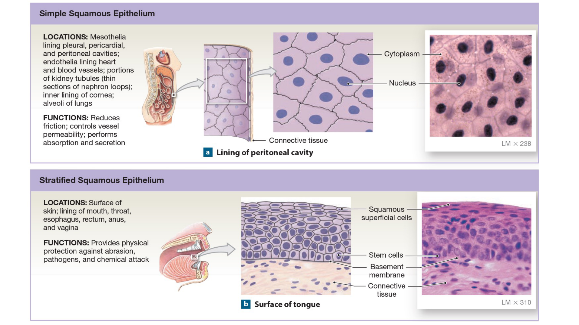
| Simple Squamous | Single layer of flat, scale-like cells with disc-shaped nuclei | Lung alveoli (gas exchange), kidney glomeruli Bowman's capsule (filtration), endothelium (lining blood/lymph vessels), mesothelium (pericardium, pleura, peritoneum). |
| Simple Cuboidal | Single layer of cube-shaped cells with central round spherical nuclei | Kidney tubules (secretion/absorption), surface of ovary, thyroid gland follicles. |
| Simple Columnar | Single layer of tall rectangular cells with oval basal nuclei; often with microvilli or cilia, and goblet cells | Non-ciliated: GI tract lining (stomach, small & large intestine, gallbladder; absorption/secretion). Ciliated: Uterine (Fallopian) tubes (propels oocyte). |
| Pseudostratified Columnar | Single layer appearing multi-layered; all cells touch basement membrane but not all reach surface; ciliated with goblet cells | Lining of upper respiratory tract: nasal cavity, trachea, bronchi (mucus secretion and ciliary escalation of debris). |
| Stratified Squamous (Keratinized) | Multiple layers; superficial cells are dead, anucleate, filled with water-resistant keratin | Epidermis of skin (protection against abrasion, pathogens, desiccation). |
| Stratified Squamous (Non-keratinized) | Multiple layers; superficial cells living, nucleated, moist | Oral cavity, pharynx, esophagus, vagina, anal canal (protection against mechanical abrasion in moist cavities). |
| Transitional (Urothelium) | Stratified with dome-shaped/umbrella apical cells that flatten when stretched | Urinary bladder, ureters, renal calyces, proximal urethra (permits reversible distension while maintaining impermeable urine barrier). |
เนื้อเยื่อบุผิวจำแนกตามจำนวนชั้น (Simple = ชั้นเดียว, Stratified = หลายชั้น, Pseudostratified = หลายชั้นเทียม) และรูปร่างเซลล์ชั้นบนสุด (Squamous = แบน, Cuboidal = ลูกบาศก์, Columnar = ทรงสูง, Transitional = เปลี่ยนรูปร่างได้):
| ชนิดของเนื้อเยื่อบุผิว | ลักษณะทางมิญชวิทยา | ตำแหน่งที่พบและหน้าที่สำคัญ |
|---|---|---|
| Simple Squamous | เซลล์แบนชั้นเดียว นิวเคลียสแบน | ถุงลมปอด (แลกเปลี่ยนก๊าซ), โกลเมอรูลัสในไต (กรองของเสีย), Endothelium (เยื่อบุด้านในหลอดเลือดและหัวใจ), Mesothelium (เยื่อหุ้มปอด หัวใจ ช่องท้อง) |
| Simple Cuboidal | เซลล์ทรงลูกบาศก์ชั้นเดียว นิวเคลียสกลมอยู่ตรงกลาง | ท่อไต (ดูดซึมและขับสาร), ผิวรังไข่, ต่อมไทรอยด์ |
| Simple Columnar | เซลล์ทรงกระบอกสูงชั้นเดียว นิวเคลียสรูปไข่อยู่ใกล้ฐานเซลล์ มี Microvilli และ Goblet cells | ชนิดไม่มีขน: ทางเดินอาหาร (กระเพาะอาหาร, ลำไส้เล็ก, ลำไส้ใหญ่, ถุงน้ำดี; ดูดซึมและหลั่งเมือก). ชนิดมี Cilia: ท่อนำไข่ (พัดโบกเซลล์ไข่) |
| Pseudostratified Ciliated Columnar | เซลล์ชั้นเดียวแต่นิวเคลียสอยู่หลายระดับทำให้ดูเหมือนหลายชั้น เซลล์ทุกเซลล์แตะ Basement membrane มีขนกวัด (Cilia) และ Goblet cells | ทางเดินหายใจส่วนบน (โพรงจมูก, หลอดลมคอ Trachea, หลอดลม Bronchi; ดักจับฝุ่นละอองและพัดโบกเสมหะ) |
| Stratified Squamous (Keratinized) | เซลล์แบนหลายชั้น ชั้นบนสุดเป็นเซลล์ตาย ไม่มีนิวเคลียส อัดแน่นด้วยเคราติน | ผิวหนังชั้นนอกสุด (Epidermis; ป้องกันการเสียดสี การสูญเสียน้ำ และเชื้อโรค) |
| Stratified Squamous (Non-keratinized) | เซลล์แบนหลายชั้น ชั้นบนสุดยังมีชีวิต มีนิวเคลียส ผิวชุ่มชื้น | ช่องปาก, คอหอย, หลอดอาหาร, ช่องคลอด, ทวารหนัก (ทนการเสียดสีในบริเวณที่ชื้น) |
| Transitional Epithelium (Urothelium) | เซลล์หลายชั้น เซลล์ชั้นบนมีลักษณะโค้งมนคล้ายร่ม (Umbrella / Dome cell) สามารถยืดแบนได้เมื่อมีปัสสาวะเต็ม | กระเพาะปัสสาวะ, ท่อไต (Ureter), กรวยไต (ยืดขยายได้โดยไม่ฉีกขาดและไม่รั่วซึม) |
3. Connective Tissue Classification & Components / 3. เนื้อเยื่อเกี่ยวพัน องค์ประกอบ และการจำแนกชนิด
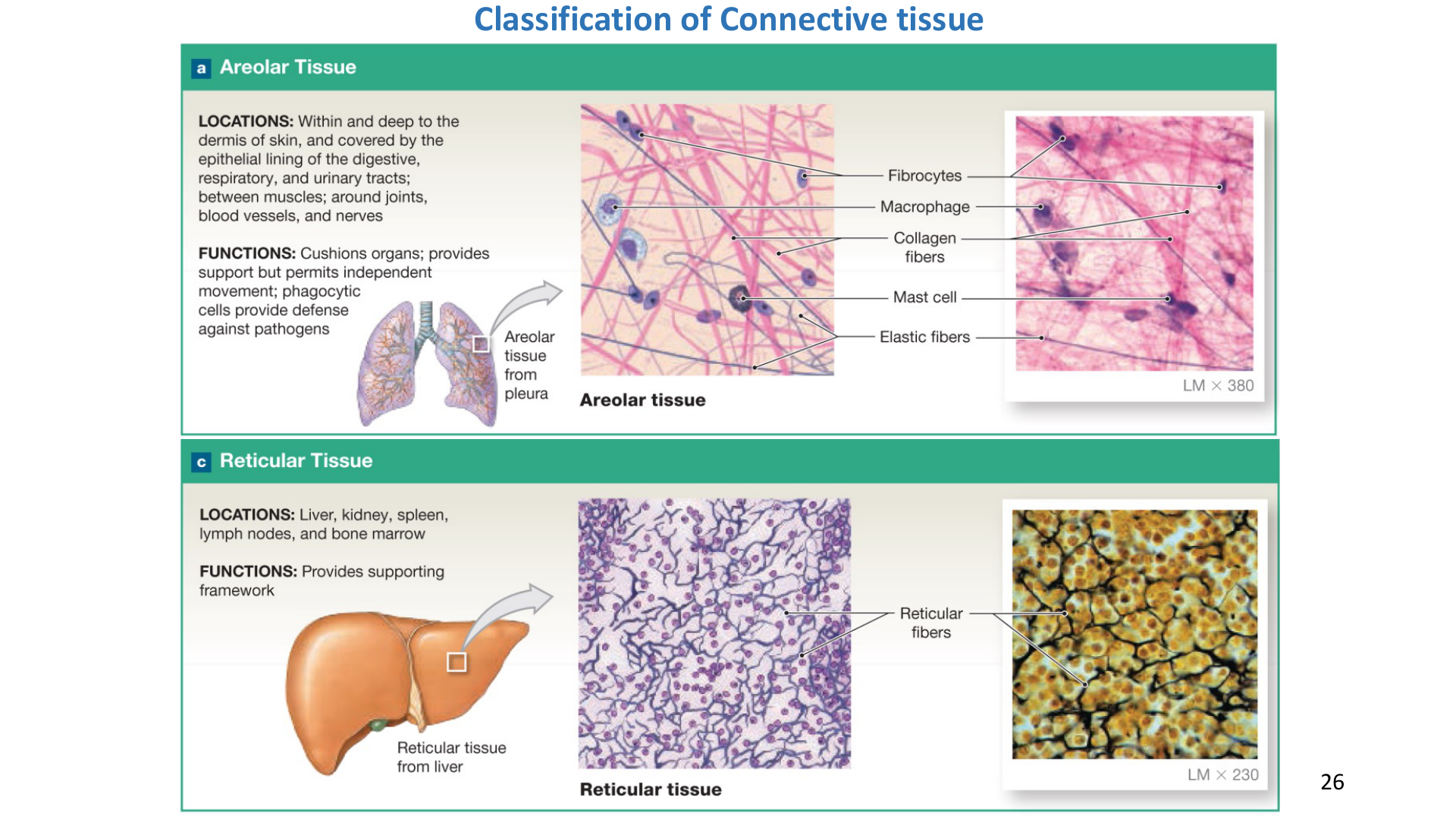
Connective tissue consists of cells widely dispersed in an Extracellular Matrix (ECM):
- Matrix Components:
- Ground substance: Gel-like medium of glycosaminoglycans (hyaluronic acid, chondroitin sulfate) and proteoglycans.
- Fibers: Collagen fibers (thick, resists tensile pulling), Elastic fibers (elastin/fibrillin, stretch and recoil up to 150%), Reticular fibers (delicate Type III collagen meshwork supporting soft organ stroma).
- Cell Types: Fibroblasts (synthesize fibers/ground substance), Adipocytes (fat storage), Macrophages (phagocytosis), Plasma cells (antibodies), Mast cells (histamine, heparin in inflammation), White blood cells.
Connective Tissue Classification
- Loose Connective Tissue:
- Areolar: Loose meshwork; lamina propria under epithelia, wraps organs and nerves.
- Adipose: Dominated by adipocytes; thermal insulation, energy depot, organ cushion.
- Reticular: Stroma of lymphoid organs (spleen, lymph nodes, bone marrow).
- Dense Connective Tissue:
- Dense Regular: Parallel, dense collagen bundles; withstands high unidirectional tension (tendons, ligaments, aponeuroses).
- Dense Irregular: Randomly interwoven collagen bundles; withstands multidirectional pull (reticular layer of dermis, periosteum, organ capsules).
- Elastic: Abundant parallel elastic fibers; permits expansion and recoil (aorta wall, vocal cords, ligamentum flavum).
- Specialized Connective Tissues: Cartilage (Hyaline, Elastic, Fibrocartilage), Bone (Compact, Spongy), and Fluid (Blood and Lymph).
เนื้อเยื่อเกี่ยวพันประกอบด้วยเซลล์ที่ล้อมรอบด้วย สารเคลือบเซลล์ (Extracellular Matrix - ECM):
- องค์ประกอบของ Matrix:
- Ground substance: สารกึ่งเหลวกึ่งเจล ประกอบด้วยน้ำ สารไกลโคสะมิโนไกลแคน (Hyaluronic acid, Chondroitin sulfate)
- เส้นใย (Fibers): Collagen fibers (หนา เหนียว แข็งแรง ทนแรงดึง), Elastic fibers (ยืดหยุ่นได้ 150% และดีดกลับได้), Reticular fibers (เส้นใยร่างแหละเอียด Type III collagen ค้ำจุนอวัยวะ)
- ชนิดของเซลล์: ไฟโบรบลาสต์ (Fibroblast = สร้างเส้นใยและ matrix), เซลล์ไขมัน (Adipocyte), แมคโครฟาจ (กลืนกินเชื้อโรค), พลาสมาเซลล์ (สร้างแอนติบอดี), แมสต์เซลล์ (หลั่งฮีสตามีนและเฮพาริน)
การจำแนกเนื้อเยื่อเกี่ยวพัน
- เนื้อเยื่อเกี่ยวพันชนิดหลวม (Loose Connective Tissue):
- Areolar: เส้นใยสานโปร่ง อยู่ใต้เนื้อเยื่อบุผิว (Lamina propria) หุ้มหลอดเลือดและเส้นประสาท
- Adipose: เซลล์ไขมันอัดแน่น ป้องกันความร้อน เป็นแหล่งพลังงาน และกันกระแทก
- Reticular: โครงร่างแหในอวัยวะน้ำเหลือง (ม้าม, ต่อมน้ำเหลือง, ไขกระดูก)
- เนื้อเยื่อเกี่ยวพันชนิดแน่นทึบ (Dense Connective Tissue):
- Dense Regular: เส้นใยคอลลาเจนเรียงขนานกันอย่างเป็นระเบียบ ทนแรงดึงในทิศทางเดียวได้ดีเยี่ยม (เอ็นกล้ามเนื้อ Tendon, เอ็นยึดข้อ Ligament)
- Dense Irregular: เส้นใยคอลลาเจนสานกันแน่นไม่เป็นระเบียบ ทนแรงดึงได้หลายทิศทาง (ผิวหนังชั้นหนังแท้ Reticular layer, เยื่อหุ้มกระดูก Periosteum)
- Elastic: เส้นใยอิลาสติกหนาแน่น ยืดหยุ่นและคืนตัวได้ดี (ผนังหลอดเลือดแดงใหญ่ Aorta, เส้นเสียง Vocal cord)
- เนื้อเยื่อเกี่ยวพันชนิดพิเศษ: กระดูกอ่อน (Cartilage), กระดูกแข็ง (Bone), และเนื้อเยื่อเกี่ยวพันชนิดของเหลว (เลือดและน้ำเหลือง)
4. Integumentary System (Skin & Strata) / 4. ระบบผิวหนัง ชั้นของหนังกำพร้า หนังแท้ และตัวรับความรู้สึก
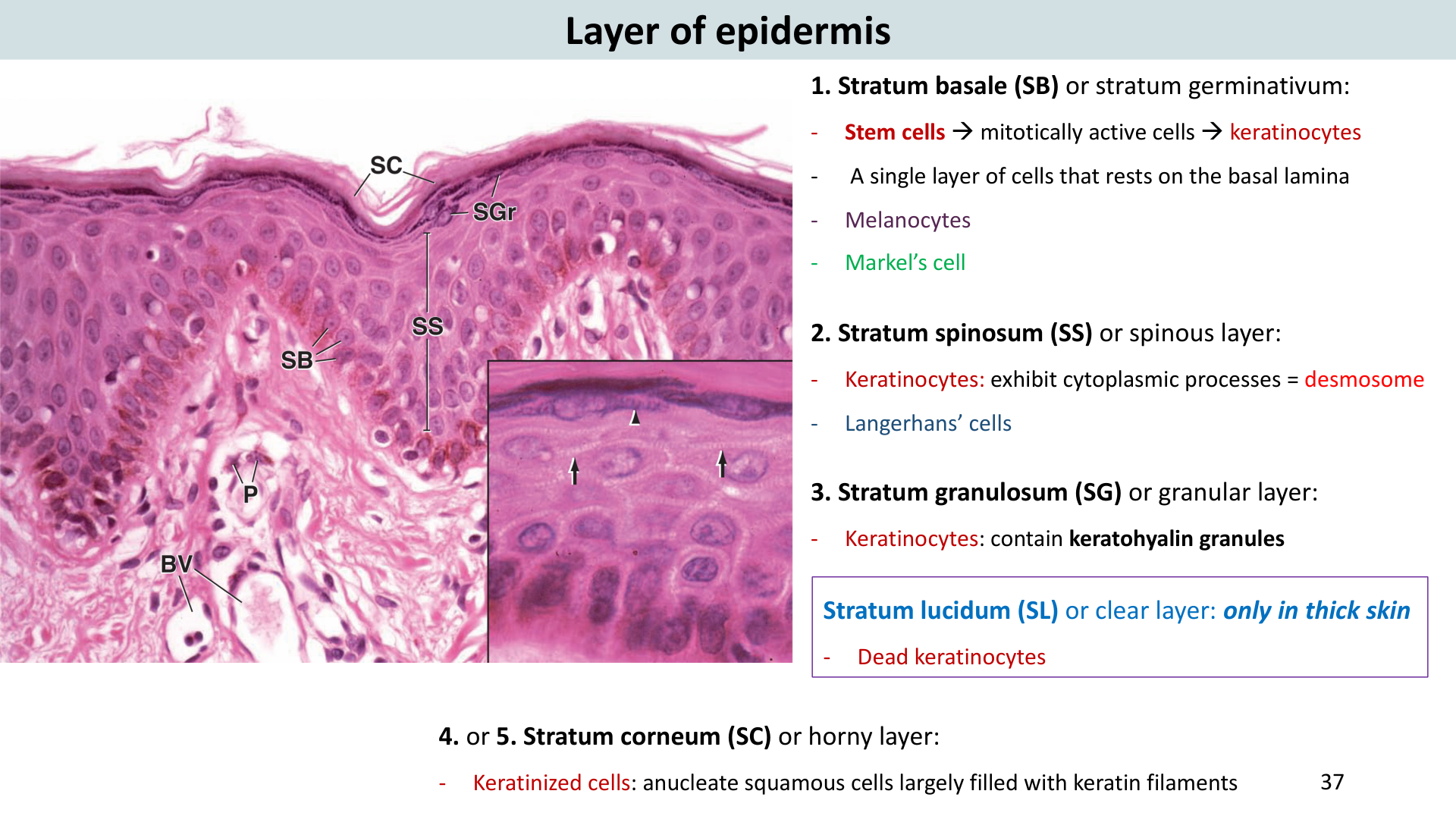
The skin (integument) consists of two main anatomical layers resting on a subcutaneous base:
1. Epidermis (Avascular keratinized stratified squamous)
5 Strata from deep to superficial (mnemonic: "Before Signing, Get Legal Counsel"):
- Stratum Basale (Germinativum): Single row of stem cells on basal lamina; highly mitotic; contains melanocytes (produce UV-protective melanin) and Merkel cells (touch).
- Stratum Spinosum: 8–10 layers of cells connected by prominent desmosomes; contains Langerhans cells (dendritic antigen-presenting immune cells).
- Stratum Granulosum: 3–5 layers; cells undergoing apoptosis; contain keratohyalin and lamellar lipid granules (waterproofing).
- Stratum Lucidum: Clear layer of dead flat cells; present ONLY in thick skin (palms and soles).
- Stratum Corneum: 20–30 layers of dead, anucleate, keratin-packed scales continually shed (desquamation).
2. Dermis (Vascular, innervated dense irregular CT)
- Papillary Layer (superficial 20%): Areolar CT; dermal papillae protruding into epidermal ridges (fingerprints); houses capillary loops, Meissner's corpuscles (light touch), and free nerve endings.
- Reticular Layer (deep 80%): Dense irregular CT with coarse collagen; contains hair roots, sebaceous glands, sweat glands, and deep Pacinian corpuscles (vibration and deep pressure).
3. Hypodermis (Subcutis / Superficial Fascia)
Adipose and loose connective tissue; anchors skin to underlying deep fascia; provides thermal insulation and mechanical shock absorption.
Cutaneous Sensory Receptors
- Free nerve endings: Pain (nociceptors), temperature (cold/warmth), itch.
- Meissner's (tactile) corpuscles: Light discriminative touch, low-frequency vibration (in dermal papillae).
- Pacinian (lamellar) corpuscles: Deep pressure, high-frequency vibration (deep dermis/hypodermis).
- Ruffini corpuscles: Skin stretch, continuous pressure.
ผิวหนัง (Skin / Integument) ประกอบด้วย 2 ชั้นหลัก วางอยู่บนชั้นเนื้อเยื่อใต้ผิวหนัง:
1. หนังกำพร้า (Epidermis: เซลล์บุผิว Stratified squamous ชนิด Keratinized)
แบ่งออกเป็น 5 ชั้นย่อย เรียงจากชั้นลึกสุดขึ้นสู่ผิวบนสุด:
- Stratum Basale (Germinativum): ชั้นลึกสุด เซลล์แถวเดียวแบ่งตัวตลอดเวลา (Mitosis) มีเซลล์สร้างเม็ดสี Melanocyte และเซลล์รับสัมผัส Merkel cell
- Stratum Spinosum: เซลล์ 8-10 ชั้น เชื่อมกันด้วย Desmosome แน่นหนา มีเซลล์ภูมิคุ้มกัน Langerhans cell คอยดักจับเชื้อโรค
- Stratum Granulosum: 3-5 ชั้น เซลล์เริ่มตาย สะสมเม็ด Keratohyalin และ Lamellar granules ที่ปล่อยไขมันกันน้ำ
- Stratum Lucidum: ชั้นใส เซลล์ตายแล้ว แบน ไม่มีนิวเคลียส พบเฉพาะในผิวหนังหนา (Thick skin: ฝ่ามือและฝ่าเท้า) เท่านั้น
- Stratum Corneum: ชั้นนอกสุด เซลล์ตาย 20-30 ชั้น อัดแน่นด้วยเคราติน ลอกหลุดเป็นขี้ไคลตลอดเวลา
2. หนังแท้ (Dermis: มีหลอดเลือดและเส้นประสาทหนาแน่น)
- Papillary Layer (ชั้นบน 20%): เนื้อเยื่อ Areolar ยื่นขึ้นไปเป็นปุ่ม Dermal papillae (ทำให้เกิดลายนิ้วมือ) มีหลอดเลือดฝอยเลี้ยงหนังกำพร้า และ Meissner's corpuscle รับสัมผัสแผ่วเบา
- Reticular Layer (ชั้นล่าง 80%): เนื้อเยื่อ Dense irregular CT เหนียวทนทาน มีรูขุมขน ต่อมไขมัน ต่อมเหงื่อ และ Pacinian corpuscle รับแรงกดลึกและการสั่นสะเทือน
3. ชั้นใต้ผิวหนัง (Hypodermis / Subcutaneous tissue)
เนื้อเยื่อไขมันและเนื้อเยื่อเกี่ยวพันหลวม ช่วยกักเก็บพลังงาน กันกระแทก กันความร้อน และยึดผิวหนังเข้ากับพังผืดกล้ามเนื้อด้านล่าง
ตัวรับความรู้สึกที่ผิวหนัง (Cutaneous Receptors)
- Free nerve endings: ปลายประสาทอิสระ รับความเจ็บปวด (Pain) และอุณหภูมิ (ร้อน/เย็น)
- Meissner's corpuscles: รับสัมผัสแผ่วเบา (Light touch) อยู่ที่ Dermal papillae
- Pacinian corpuscles: รับแรงกดลึก (Deep pressure) และการสั่นสะเทือนความถี่สูง (Vibration) อยู่ชั้นลึก
- Ruffini corpuscles: รับแรงดึงยืดของผิวหนัง (Stretch)
5. Burns & Wound Healing Phases / 5. แผลไฟไหม้ (Burns) และ 4 ระยะของการสมานแผล (Wound Healing)
Classification of Burns
- First-degree burn: Damage confined to epidermis (erythema, pain, edema, no blisters; e.g. mild sunburn). Heals in 3–6 days.
- Second-degree burn: Destroys epidermis and upper papillary/reticular dermis (blisters, severe pain, edema; partial-thickness). Heals in 2–4 weeks.
- Third-degree burn: Destroys entire thickness of skin into hypodermis (charred/pearly white, painless due to sensory nerve ending destruction; full-thickness). Requires skin grafting.
4 Phases of Deep Wound Healing
- Hemostasis (Clotting): Immediate vasoconstriction, platelet plug aggregation, and fibrin mesh clot formation to arrest bleeding.
- Inflammatory Phase: Vasodilation and increased vascular permeability; infiltration of neutrophils followed by monocytes (macrophages) to phagocytose pathogens, debris, and necrotic tissue. Cardinal signs: rubor (redness), calor (heat), tumor (swelling), dolor (pain).
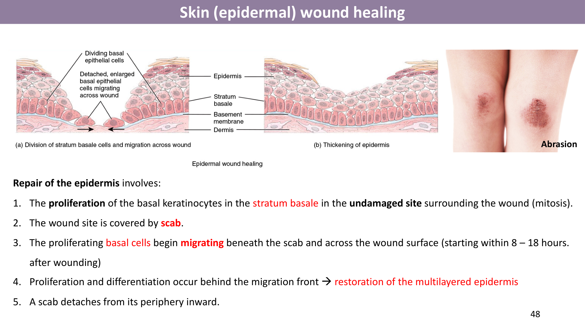
- Proliferative Phase: Re-epithelialization beneath scab; fibroblasts synthesize abundant collagen and ground substance; angiogenesis (new capillary buds) forms vascularized granulation tissue; wound contraction by myofibroblasts.
- Maturation (Remodeling) Phase: Scab sloughs off; collagen fibers reorganized and cross-linked along lines of tension; capillaries recede; scar tissue formation (fibrosis). Complications: Hypertrophic scar (confined within wound borders) vs. Keloid (collagen overgrowth extending beyond original boundaries).
การจำแนกระดับของแผลไฟไหม้ (Burns)
- First-degree burn (ระดับที่ 1): เสียหายเฉพาะชั้นหนังกำพร้า ผิวแดง ปวด บวม ไม่มีตุ่มน้ำใส (เช่น ผิวไหม้แดด) หายเองใน 3-6 วัน
- Second-degree burn (ระดับที่ 2): เสียหายถึงชั้นหนังแท้ เกิดตุ่มน้ำใส (Blister) ปวดแสบปวดร้อนมาก บวม หายใน 2-4 สัปดาห์
- Third-degree burn (ระดับที่ 3): ทำลายผิวหนังทุกชั้นทะลุถึงชั้นใต้ผิวหนัง ผิวมีสีขาวซีดหรือไหม้เกรียม ไม่มีความเจ็บปวดเนื่องจากปลายประสาทถูกทำลายหมด ต้องปลูกถ่ายผิวหนัง
4 ระยะของการสมานแผลลึก (Deep Wound Healing)
- Hemostasis (ระยะห้ามเลือด): เส้นเลือดหดตัวทันที เกล็ดเลือดเกาะกลุ่ม และเกิดตาข่ายไฟบรินเป็นลิ่มเลือดหยุดการไหลของเลือด
- Inflammatory Phase (ระยะการอักเสบ): หลอดเลือดขยายตัว นิวโทรฟิลและแมคโครฟาจเข้าสู่บาดแผลเพื่อกลืนกินเชื้อโรคและเศษเนื้อเยื่อตาย อาการแสดง 4 ประการ: ปวด บวม แดง ร้อน
- Proliferative Phase (ระยะการสร้างเนื้อเยื่อ): เซลล์เยื่อบุผิวแบ่งตัวคลุมแผลใต้สะเก็ด ไฟโบรบลาสต์สร้างคอลลาเจน มีการงอกของหลอดเลือดใหม่เกิดเป็น Granulation tissue (เนื้อเยื่อแกรนูเลชันสีชมพูแดง) และดึงรั้งปากแผลให้แคบลง
- Maturation / Remodeling Phase (ระยะการจัดระเบียบเนื้อเยื่อ): สะเก็ดแผลหลุด คอลลาเจนจัดเรียงตัวเป็นระเบียบตามแนวแรงตึง เกิดเป็นแผลเป็น (Scar / Fibrosis) หากสร้างคอลลาเจนมากเกินไปอาจเกิด แผลเป็นนูนคีลอยด์ (Keloid) ที่ลุกลามออกนอกขอบเขตแผลเดิม