Topic 2: Introduction to Physiology / บทที่ 2: บทนำสรีรวิทยาและการขนส่งผ่านเยื่อหุ้มเซลล์
Homeostasis, cell membrane structure, passive and active transport mechanisms, osmolarity vs tonicity, and resting membrane potential (RMP).
ภาวะธำรงดุล โครงสร้างเยื่อหุ้มเซลล์ กลไกการขนส่งแบบพาสซีฟและแอกทีฟ ความต่างของออสโมลาริตีกับโทนิซิตี และศักย์เยื่อเซลล์ระยะพัก (RMP)
1. Homeostasis & Core Principles / 1. ภาวะธำรงดุลและหลักการสรีรวิทยา
Physiology is the study of how living organisms function, focusing on cause-and-effect physical and chemical mechanisms.
- Homeostasis: Maintenance of dynamic constancy of the internal environment (ECF: body temperature, pH, blood glucose, ion concentrations) despite external changes.
- Negative Feedback Loop: Chief regulatory mechanism. A deviation from normal setpoint triggers a response in the opposite direction (e.g. insulin secretion lowering blood glucose, sweating lowering body temperature).
- Positive Feedback Loop: Rare; amplifies initial stimulus until a specific endpoint is reached (e.g. oxytocin in labor child delivery, platelet plug formation in clotting).
สรีรวิทยา (Physiology) คือการศึกษากลไกการทำงานของสิ่งมีชีวิต โดยเน้นที่กระบวนการทางเคมีและกายภาพที่เชื่อมโยงเป็นเหตุเป็นผล
- ภาวะธำรงดุล (Homeostasis): ความสามารถในการรักษาสภาวะแวดล้อมภายในร่างกาย (ECF: อุณหภูมิ, pH, ระดับน้ำตาลในเลือด, ความเข้มข้นของไอออน) ให้คงที่และเหมาะสมต่อการดำรงชีวิต
- การควบคุมแบบป้อนกลับทางลบ (Negative Feedback): กลไกหลักของร่างกาย เมื่อค่าเบี่ยงเบนไปจากจุดปกติ (set point) ร่างกายจะตอบสนองในทิศทางตรงกันข้ามเพื่อดึงกลับสู่ภาวะปกติ (เช่น การหลั่งอินซูลินลดน้ำตาล, การหลั่งเหงื่อลดอุณหภูมิ)
- การควบคุมแบบป้อนกลับทางบวก (Positive Feedback): เกิดขึ้นน้อย กระตุ้นให้การเปลี่ยนแปลงทวีความรุนแรงขึ้นจนกว่าจะบรรลุเป้าหมาย (เช่น การหลั่งออกซิโตซินขณะคลอดบุตร, การเกาะกลุ่มของเกล็ดเลือดเมื่อเกิดแผล)
2. Cell Membrane Architecture & Permeability / 2. โครงสร้างเยื่อหุ้มเซลล์และการซึมผ่าน
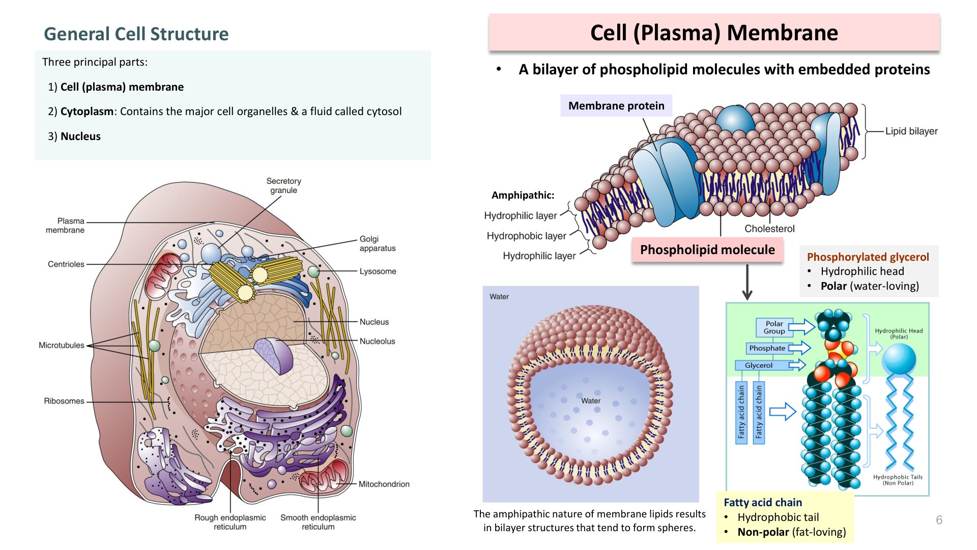
Fluid Mosaic Model: Phospholipid bilayer with embedded proteins and cholesterol:
- Phospholipids: Amphipathic; hydrophilic polar heads face outward (ECF & ICF); hydrophobic non-polar fatty acid tails sequestered in the interior.
- Cholesterol: Intercalates between fatty acid chains to regulate membrane fluidity and mechanical stability.
- Integral (Transmembrane) Proteins: Span the bilayer; serve as channels, carriers, receptors, and enzymes.
- Peripheral Proteins: Attached to outer or inner surface; anchor cytoskeleton and signal complexes.
Membrane Permeability Rules
- Freely Permeable: Small non-polar, lipid-soluble molecules (O2, CO2, N2, steroid hormones, fatty acids, fat-soluble vitamins A, D, E, K).
- Slightly Permeable: Small uncharged polar molecules (H2O, urea, glycerol).
- Impermeable (Require Transporters): Large uncharged polar molecules (glucose) and ALL charged ions (Na+, K+, Ca2+, Cl-, HCO3-, amino acids, ATP).
แบบจำลองฟลูอิดโมเสก (Fluid Mosaic Model): เยื่อหุ้มเซลล์ประกอบด้วยฟอสโฟลิพิด 2 ชั้น มีโปรตีนและคอเลสเตอรอลแทรกอยู่:
- ฟอสโฟลิพิด (Phospholipids): มีคุณสมบัติแอมฟิพาติก (Amphipathic); ส่วนหัวมีขั้วชอบน้ำ (Hydrophilic head) หันออกด้านนอกสัมผัสของเหลว ECF และ ICF; ส่วนหางไม่มีขั้วไม่ชอบน้ำ (Hydrophobic tail) หลบเข้าหากันด้านใน
- คอเลสเตอรอล (Cholesterol): แทรกอยู่ระหว่างหางกรดไขมัน ช่วยควบคุมความยืดหยุ่นและความเสถียรของเยื่อหุ้มเซลล์
- อินทิกรัลโปรตีน (Integral / Transmembrane Proteins): โปรตีนที่แทรกทะลุผ่านเยื่อหุ้มเซลล์ ทำหน้าที่เป็นช่องไอออน ตัวพา ตัวรับสัญญาณ หรือเอนไซม์
- เพอริเฟอรัลโปรตีน (Peripheral Proteins): โปรตีนที่เกาะอยู่บริเวณผิวของเยื่อหุ้มเซลล์ เชื่อมต่อกับไซโทสเกเลตันและส่งสัญญาณ
กฎการซึมผ่านของเยื่อหุ้มเซลล์
- ผ่านได้อิสระ: โมเลกุลขนาดเล็ก ไม่มีขั้ว ละลายได้ดีในไขมัน (O2, CO2, N2, สเตียรอยด์ฮอร์โมน, กรดไขมัน, วิตามิน A, D, E, K)
- ผ่านได้เล็กน้อย: โมเลกุลมีขั้วขนาดเล็กที่ไม่มีประจุ (H2O, ยูเรีย, กลีเซอรอล)
- ผ่านไม่ได้ (ต้องอาศัยโปรตีนตัวพา/ช่อง): โมเลกุลขนาดใหญ่ (กลูโคส) และ ไอออนที่มีประจุทั้งหมด (Na+, K+, Ca2+, Cl-, กรดอะมิโน, ATP)
3. Passive Transport & Osmosis vs. Tonicity / 3. การขนส่งแบบพาสซีฟ และออสโมซิสกับโทนิซิตี
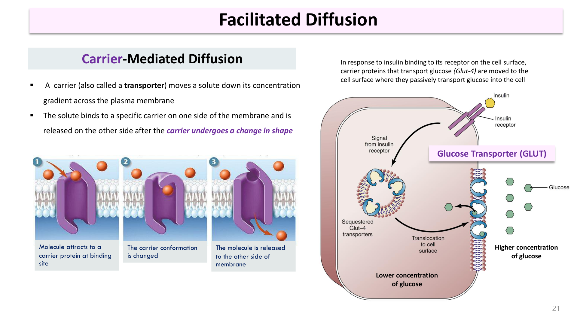
Passive Transport: Down electrochemical/concentration gradient; requires NO cellular ATP energy.
- Simple Diffusion: Movement of solutes through lipid bilayer down gradient until equilibrium.
- Channel-Mediated Diffusion: Non-gated (leak) channels (e.g. K+ leak) or Gated channels (voltage-gated, ligand-gated, mechanically-gated).
- Facilitated Diffusion: Carrier proteins undergo conformational shifts (e.g. GLUT-4 mobilized to sarcolemma by insulin).
- Osmosis: Net movement of water across a semi-permeable membrane from low solute concentration (high water) to high solute concentration (low water).
Osmolarity vs. Tonicity
- Osmolarity: Total concentration of all solute particles per liter of solution (normal plasma = ~300 mOsm/L).
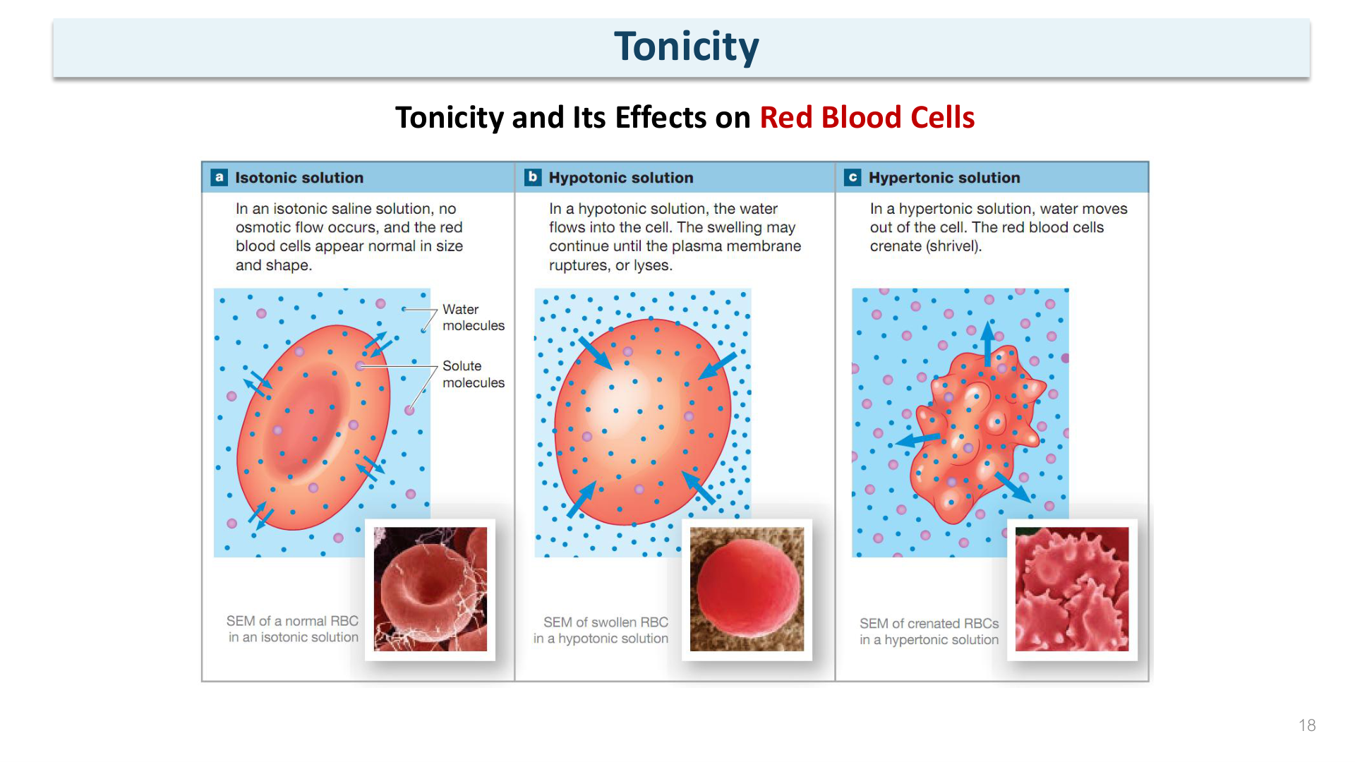
- Tonicity: Effect of solution on cell volume; determined ONLY by non-penetrating solutes:
- Isotonic (~300 mOsm/L non-penetrating): No net water flux; cell volume unchanged (e.g., 0.9% NaCl Normal Saline, 5% Dextrose D5W).
- Hypotonic (<300 mOsm/L): Water flows INTO cell ➔ cell swells and may burst (hemolysis in RBCs).
- Hypertonic (>300 mOsm/L): Water leaves cell ➔ cell shrinks (crenation; clinical use: Mannitol IV for cerebral edema).
การขนส่งแบบไม่ใช้พลังงาน (Passive Transport): เคลื่อนที่ตามความต่างของความเข้มข้น ไม่ต้องใช้พลังงาน ATP:
- การแพร่ธรรมดา (Simple Diffusion): สารละลายผ่านชั้นไขมันได้โดยตรงจากเข้มข้นมากไปน้อย
- การแพร่ผ่านช่องโปรตีน (Channel-Mediated): ช่องเปิดตลอด (Leak channel เช่น K+ leak) หรือช่องที่มีประตูปิดเปิด (Gated channels: มีทั้งประตูกระแสไฟฟ้า, ประตูสารเคมี, ประตูแรงกล)
- การแพร่แบบฟาซิลิเทต (Facilitated Diffusion): อาศัยโปรตีนตัวพาที่มีการเปลี่ยนรูป (เช่น ตัวพากลูโคส GLUT-4 ในเซลล์กล้ามเนื้อที่ถูกกระตุ้นโดยอินซูลิน)
- ออสโมซิส (Osmosis): การเคลื่อนที่สุทธิของน้ำผ่านเยื่อเลือกผ่าน จากบริเวณที่มีตัวถูกละลายต่ำ (น้ำมาก) ไปยังบริเวณที่มีตัวถูกละลายสูง (น้ำน้อย)
ความต่างระหว่าง Osmolarity กับ Tonicity
- Osmolarity (ออสโมลาริตี): ความเข้มข้นรวมของอนุภาคทั้งหมดในสารละลาย 1 ลิตร (พลาสมาปกติ = ประมาณ 300 mOsm/L)
- Tonicity (โทนิซิตี): ความสามารถของสารละลายภายนอกในการเปลี่ยนปริมาตรเซลล์ โดยคิดเฉพาะ สารที่ไม่สามารถซึมผ่านเยื่อหุ้มเซลล์ได้ (Non-penetrating solutes):
- Isotonic Solution: ความเข้มข้นเท่ากับภายในเซลล์ ไม่มีน้ำไหลเข้าออกสุทธิ เซลล์คงรูปร่างเดิม (เช่น 0.9% NSS, 5% Dextrose)
- Hypotonic Solution: ความเข้มข้นสารละลายภายนอกต่ำกว่าในเซลล์ น้ำจะออสโมซิสเข้าสู่เซลล์ ➔ เซลล์เต่งและแตกออก (Hemolysis)
- Hypertonic Solution: ความเข้มข้นภายนอกสูงกว่าในเซลล์ น้ำจะไหลออกจากเซลล์ ➔ เซลล์เหี่ยว (Crenation; เช่น การให้ Mannitol เพื่อลดภาวะสมองบวม)
4. Active Transport Mechanisms / 4. กลไกการขนส่งแบบใช้พลังงาน (Active Transport)
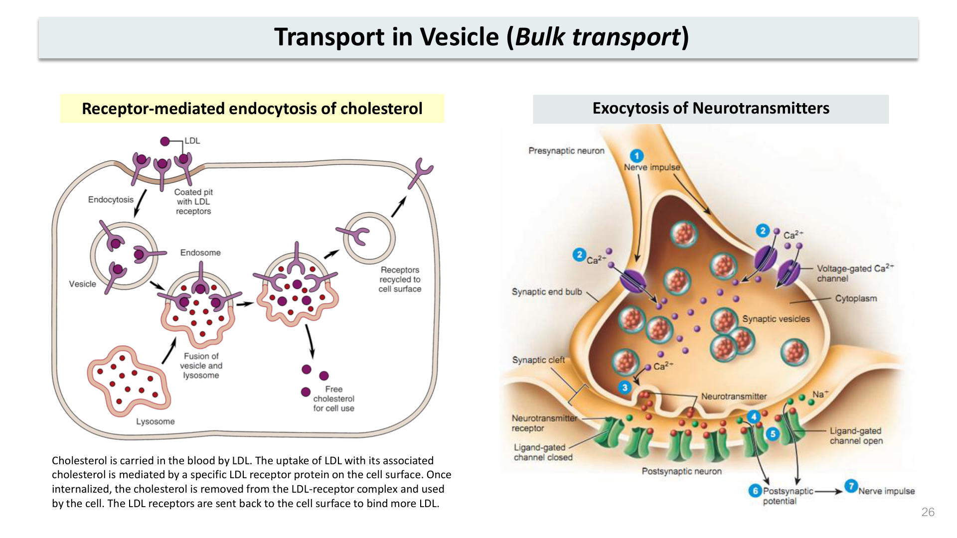
Active Transport: Moves solutes AGAINST their concentration gradient; requires cellular energy.
- Primary Active Transport: Directly hydrolyzes ATP.
- Na+/K+ ATPase Pump: Hydrolyzes 1 ATP to pump 3 Na+ OUT of cell and 2 K+ IN against gradients. Crucial for cell osmotic volume and maintaining resting membrane potential.
- Ca2+ ATPase Pump (SERCA): Pumps Ca2+ back into sarcoplasmic reticulum to end muscle contraction.
- Secondary Active Transport: Uses the potential energy stored in an ionic gradient established by primary transport (usually the Na+ gradient):
- Symport (Cotransport): Solutes move in SAME direction (e.g. SGLT transports Na+ and glucose together into intestinal/kidney epithelial cells).
- Antiport (Countertransport): Solutes move in OPPOSITE directions (e.g. Na+/H+ exchanger, Na+/Ca2+ exchanger).
- Vesicular Transport (Bulk): Endocytosis (Phagocytosis "cell eating", Pinocytosis "cell drinking", Receptor-mediated endocytosis via clathrin pits) and Exocytosis (neurotransmitter release).
การขนส่งแบบใช้พลังงาน (Active Transport): ลำเลียงสารต้านความเข้มข้น ต้องใช้พลังงานจากเซลล์:
- Primary Active Transport (ปฐมภูมิ): ใช้พลังงานจากการสลายพันธะ ATP โดยตรง
- Na+/K+ ATPase Pump: สลาย 1 ATP เพื่อปั๊ม 3 Na+ ออกนอกเซลล์ และนำ 2 K+ เข้าสู่ในเซลล์ รักษาความต่างศักย์และปริมาตรเซลล์
- Ca2+ ATPase Pump (SERCA): ปั๊ม Ca2+ กลับเข้าสู่ SR เพื่อหยุดการหดตัวของกล้ามเนื้อ
- Secondary Active Transport (ทุติยภูมิ): ไม่ใช้ ATP โดยตรง แต่ใช้พลังงานศักย์จากการไหลลงของเกรเดียนต์ไอออนที่สร้างไว้โดย Primary transport:
- Symport (ตัวพาร่วมทางเดียวกัน): ขนส่งสารไปในทิศทางเดียวกัน (เช่น SGLT ขนส่ง Na+ พร้อมกับกลูโคสเข้าสู่เซลล์เยื่อบุลำไส้และไต)
- Antiport (ตัวพาสวนทาง): ขนส่งสารในทิศทางตรงกันข้าม (เช่น Na+/Ca2+ exchanger, Na+/H+ exchanger)
- Vesicular Transport (การลำเลียงผ่านถุงเวสิเคิล): เอนโดไซโทซิส (ฟาโกไซโทซิส = กินของแข็ง, พิโนไซโทซิส = ดื่มสารละลาย, การนำเข้าอาศัยตัวรับผ่าน Clathrin) และเอกโซไซโทซิส (การหลั่งสารสื่อประสาท)
5. Resting Membrane Potential (RMP) / 5. ศักย์เยื่อเซลล์ระยะพัก (Resting Membrane Potential - RMP)
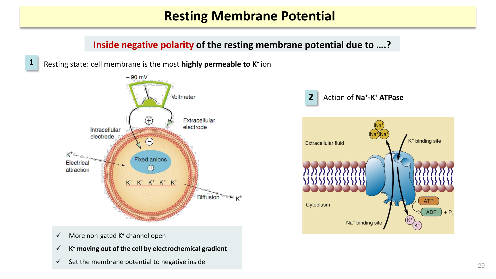
Resting Membrane Potential (RMP): Electrical voltage difference across the plasma membrane of excitable cells at rest (Em = -70 mV in neurons, -90 mV in skeletal muscle fibers). The inside of the cell is negative relative to the outside.
Two Major Mechanisms Producing Negative Inside Charge:
- High Resting Permeability to K+: The cell membrane has 50–100 times more open K+ leak channels than Na+ leak channels. K+ diffuses OUT down its steep concentration gradient, leaving behind impermeable, negatively charged intracellular proteins and organic phosphates.
- Electrogenic Na+/K+ Pump: Pumping 3 positive charges out (3 Na+) for every 2 positive charges brought in (2 K+) directly contributes -5 to -10 mV to the resting potential and prevents concentration gradient dissipation.
Membrane State Changes
- Depolarization: Membrane potential becomes less negative / moves closer to 0 mV (e.g. from -70 mV to -50 mV or +30 mV via Na+ influx).
- Repolarization: Returning toward negative resting baseline (via K+ efflux).
- Hyperpolarization: Membrane potential becomes more negative than baseline RMP (e.g. from -70 mV to -85 mV).
ศักย์เยื่อเซลล์ระยะพัก (RMP): ค่าความต่างศักย์ไฟฟ้าระหว่างภายในและภายนอกเซลล์ขณะพัก (-70 mV ในเซลล์ประสาท, -90 mV ในเซลล์กล้ามเนื้อโครงร่าง) โดยภายในเซลล์มีประจุเป็นลบเมื่อเทียบกับภายนอก
2 กลไกสำคัญที่ทำให้เกิดประจุลบภายในเซลล์:
- การซึมผ่านของ K+ ที่สูงมากในระยะพัก: เยื่อหุ้มเซลล์มี ช่องรั่วโพแทสเซียม (K+ leak channels) เปิดอยู่มากกว่าช่องโซเดียม 50-100 เท่า โพแทสเซียมจึงแพร่ออกนอกเซลล์ตามความเข้มข้น ทิ้งโปรตีนและฟอสเฟตขนาดใหญ่ที่มีประจุลบไว้ภายในเซลล์
- ปั๊ม Na+/K+ (Electrogenic pump): ปั๊ม 3 Na+ ออก แต่ดึงกลับมาเพียง 2 K+ ทำให้สูญเสียประจุบวกสุทธิ ช่วยสร้างประจุลบเพิ่มอีก -5 ถึง -10 mV และรักษาเกรเดียนต์ไว้ตลอดเวลา
การเปลี่ยนแปลงของศักย์เยื่อเซลล์
- Depolarization (ดีโพลาไรเซชัน): ความต่างศักย์ภายในเซลล์มีความเป็นลบน้อยลง / เป็นบวกมากขึ้น (เช่น จาก -70 mV เป็น 0 หรือ +30 mV จากการไหลเข้าของ Na+)
- Repolarization (รีโพลาไรเซชัน): ศักย์เยื่อเซลล์ลดลงกลับสู่ค่าระยะพัก (จากการไหลออกของ K+)
- Hyperpolarization (ไฮเปอร์โพลาไรเซชัน): ภายในเซลล์เป็นลบมากกว่าค่าระยะพักปกติ (เช่น ลดลงไปถึง -85 mV)